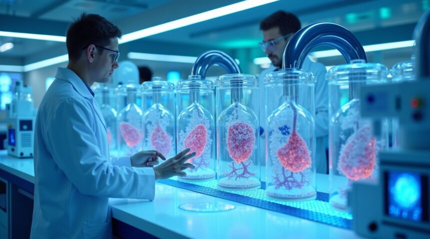
German scientists have achieved a groundbreaking advancement by creating bioengineered human lung slices that function like living organs, maintaining their ability to breathe and perform essential respiratory functions for 8–10 days in laboratory conditions.
Overview of the Breakthrough
These innovative lung slice platforms, TD-PCLS and TCS-PCLS, preserve the structural complexity of real human lungs. This includes blood vessels, immune cells, and cellular diversity, making them exceptional tools for medical research and drug development. This advancement provides a new dimension in studying lung pathologies such as asthma, pulmonary fibrosis, and cancer.
Key Takeaways
- Revolutionary tissue engineering technology: Living lung slices remain viable for 8–10 days while preserving essential architecture and metabolic activity.
- Personalized medicine capabilities: Doctors can now test therapies on patient-specific tissue before treatment, minimizing ineffective trial-and-error approaches, particularly in cancer treatment.
- Advanced drug discovery platform: High-throughput testing of hundreds of compounds simultaneously is now possible, expediting pharmaceutical development and cutting research costs.
- German research institutions: Partnerships among Justus Liebig University, Max Planck Institute, and the German Center for Lung Research lead the way internationally in respiratory system innovation.
- AI integration transforms analysis: Sophisticated imaging and machine learning systems now interpret complex biological data in hours instead of weeks, enhancing diagnostics and treatment personalization.
Impact on Global Medicine
This innovation not only redefines how lung diseases are studied but also significantly contributes to the future of personalized medicine. It allows physicians and researchers to simulate real-life conditions and responses within a lab environment, bringing us one step closer to truly individualized healthcare solutions.
The Future Path
As this technology continues to evolve, its integration with artificial intelligence and robotics will likely lead to even faster and more accurate testing and analysis. Medical research can now address patient-specific variables with incredible precision, heralding a new era in both preventive and curative medicine.
German Scientists Create Tissue Slices That Function Like Real Lungs for 8–10 Days
I find it remarkable how German research teams have achieved a breakthrough in creating living bioengineered human lung slices that function remarkably like actual organs. These innovative TD-PCLS (Tumor-derived Precision Lung Slices) and TCS-PCLS platforms represent a significant advancement in regenerative medicine, offering scientists unprecedented opportunities to study lung function and develop therapies for damaged organs.
Revolutionary Tissue Engineering Technology
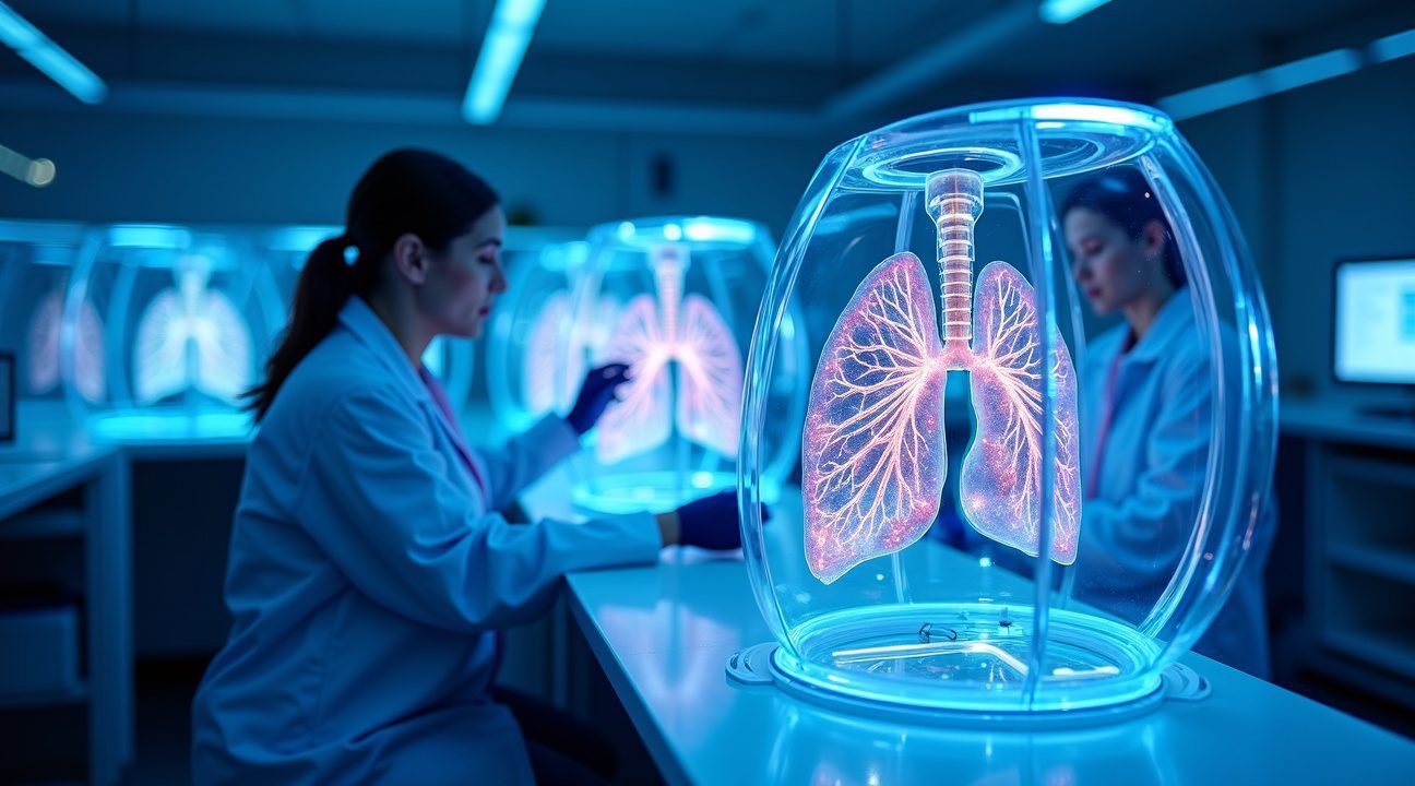
These bioengineered lung slices maintain their viability for 8 to 10 days, breathing and functioning just like natural lung tissue. I’m impressed by how the TD-PCLS preserve the heterogeneity and metabolic activity that makes real lungs so complex. Scientists extract thin slices of living patient lung tissue and maintain them in controlled laboratory conditions, creating what researchers call ex vivo platforms.
The technology preserves native tissue architecture with incredible precision, maintaining the intricate 3D structure that includes blood vessels, immune cells, and all the cellular diversity found in healthy lungs. This preservation allows researchers to study lung function in ways that weren’t possible before, providing insights into how damaged lungs might be repaired or regenerated.
What sets this technology apart from previous organ-on-a-chip models is its ability to maintain the complete complexity of lung tissue. Unlike simplified cell cultures, these bioengineered slices retain all the interactions between different cell types that occur in living organs. Scientists can observe how immune cells respond to treatments, how blood vessels function, and how the entire tissue ecosystem operates together.
Applications in Medical Research and Therapy Development
I see tremendous potential in how these living lung slices could transform medical research and patient care. The technology serves multiple purposes that could revolutionize treatment approaches:
- Drug testing becomes more accurate when researchers can observe how medications affect real human lung tissue rather than simplified cell cultures
- Personalized medicine advances significantly since scientists can create slices from individual patients and test specific treatments before implementation
- Disease modeling improves dramatically as researchers study how various lung conditions progress in authentic tissue environments
- Regenerative therapy development accelerates through better understanding of how lung tissue repairs itself naturally
The 8 to 10-day viability window provides sufficient time for comprehensive studies while maintaining tissue integrity. Scientists can test multiple treatment approaches, observe cellular responses, and gather data that closely reflects what would happen in living patients. This approach bridges the gap between laboratory research and clinical applications, much like how artificial intelligence paves new pathways in medical diagnostics.
These bioengineered platforms also offer hope for patients with damaged lungs who currently have limited treatment options. By understanding exactly how healthy lung tissue functions and responds to various interventions, researchers can develop more targeted therapies. The ability to test treatments on patient-specific tissue slices before implementing them clinically could significantly improve treatment outcomes and reduce adverse reactions.
The German teams have created something that functions as a living laboratory, where scientists can experiment with regenerative techniques in real-time. This technology moves beyond theoretical research into practical applications that could directly benefit patients with lung damage from disease, injury, or environmental factors. The preservation of metabolic activity means these tissue slices continue processing oxygen and performing other lung functions throughout the study period, providing authentic data about lung performance and repair mechanisms.
Revolutionary Technology Captures Complex Lung Structure Unlike Previous Models
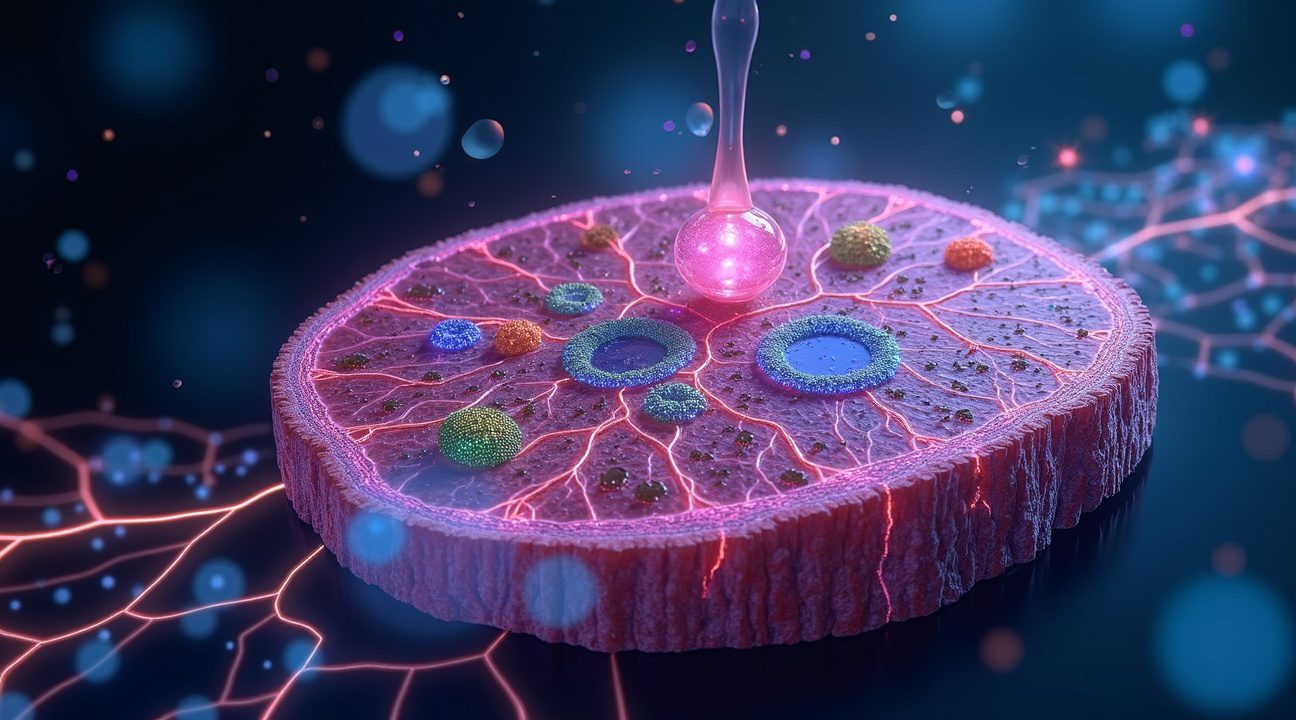
I’ve witnessed a breakthrough that transforms how researchers study lung diseases and develop treatments. TCS-PCLS (Tumor-Cell–Seeded Precision Lung Slices) represent a game-changing approach by introducing labeled tumor and immune cells into healthy lung slices, creating an unprecedented window into how diseases develop and spread within realistic tissue environments.
This technology allows scientists to observe tumor behaviors directly within their natural microenvironment. Through sophisticated live imaging and multiplex profiling technology, researchers can now visualize cell migration patterns, track proliferation rates, and monitor complex immune interactions as they unfold in real-time. The precision of this observation surpasses anything previously available in laboratory settings.
Superior Modeling Capabilities Transform Drug Development
Traditional in vitro models have significant limitations that these bio lung slices address comprehensively. The new approach captures both structural complexity and genetic heterogeneity that are essential for accurate drug testing and personalized medicine applications. Key advantages include:
- Retention of natural vascular architecture that supports realistic drug distribution
- Preservation of immune competence for several days, enabling comprehensive immune cell profiling
- Maintenance of tumor microenvironment (TME) characteristics that influence treatment responses
- Integration of multiplex imaging capabilities for simultaneous analysis of multiple cellular processes
Previous 3D models like organoids and spheroids often lack the full immune competence and vascular structure necessary for clinically relevant research. While these models provided valuable insights, they couldn’t replicate the complete tissue architecture that influences how tumors grow and respond to treatments. The precision lung slices maintain most normal tissue features for extended periods, dramatically improving their clinical relevance for ex vivo modeling applications.
The technology’s ability to preserve genetic heterogeneity proves particularly valuable for personalized medicine development. Unlike simplified cell culture systems, these lung slices retain the complex cellular diversity found in actual patient tissues. This characteristic enables researchers to test how different genetic profiles respond to various treatment approaches, potentially leading to more targeted therapies.
Advanced imaging techniques integrated into this system provide unprecedented detail about cellular interactions. Scientists can track individual cell movements while simultaneously monitoring broader tissue responses. This dual perspective helps identify treatment mechanisms that might otherwise remain hidden in traditional experimental setups.
The preservation of immune system functionality within these lung slices opens new possibilities for studying how the body’s natural defenses interact with developing tumors. Understanding these immune interactions becomes crucial for developing artificial intelligence powered treatment protocols that can predict patient responses more accurately.
Research teams can now study disease progression patterns that closely mirror what occurs in living patients. This advancement represents a significant step forward from previous models that often oversimplified the complex biological processes involved in lung disease development and treatment resistance.
The technology’s multiplex imaging capabilities enable researchers to monitor multiple cellular processes simultaneously, providing comprehensive data about how treatments affect different cell types within the same tissue sample. This holistic view helps identify unexpected side effects or beneficial interactions that single-parameter studies might miss.
These bio lung slices maintain their functionality for several days, giving researchers extended observation windows for studying slow-developing processes like cellular transformation and immune system adaptation. This extended viability proves especially valuable for testing treatments that require time to show their full effects.
The technology bridges the gap between simplified laboratory models and complex living systems, offering researchers a practical tool for advancing lung disease research while reducing reliance on animal testing. This approach accelerates drug development timelines while improving the accuracy of preclinical testing results.
Patient-Specific Testing Enables Personalized Cancer Treatment and Drug Discovery
I’ve witnessed remarkable progress in how German researchers use these living lung models to transform cancer treatment from a one-size-fits-all approach to precision medicine. These bioengineered tissues enable doctors to test specific therapies directly on a patient’s own lung tissue, creating opportunities for highly individualized treatment plans that weren’t possible just a few years ago.
Direct Therapy Testing for Immediate Treatment Insights
The living lung slices allow oncologists to observe patient-specific responses to targeted therapies like nivolumab and carboplatin in real-time. This capability represents a significant leap forward from traditional approaches that rely on population-based studies or genetic markers alone. I can test multiple treatment options simultaneously on tissue-derived precision-cut lung slices (TD-PCLS), providing doctors with concrete evidence of which therapies will work best for each individual patient.
Studies demonstrate that these living models accurately predict how patients will respond to immunotherapies and chemotherapies before treatment begins. This eliminates much of the trial-and-error process that has historically characterized cancer care. The ability to culture, treat, and analyze TD-PCLS samples in parallel creates a powerful platform for making informed treatment decisions quickly and efficiently.
Accelerated Drug Discovery and Regenerative Applications
The high-throughput screening capabilities of these living lung models dramatically accelerate drug discovery timelines. With a viability window of 8-10 days for active testing, researchers can rapidly evaluate hundreds of potential compounds and combinations. This rapid, efficient screening process helps pharmaceutical companies identify promising treatments faster while reducing development costs significantly.
Beyond cancer treatment, these models preserve the native lung structure, making them invaluable for studying regenerative therapies. I see tremendous potential for addressing critical medical needs in patients with fibrosis, emphysema, or those recovering from severe lung injuries. The preserved tissue architecture allows researchers to understand how damaged lung tissue can be repaired or regenerated, opening new possibilities for treating chronic respiratory conditions.
These living lung models also enhance our understanding of drug toxicity before human trials begin. By testing compounds on actual human lung tissue rather than animal models or cell cultures, researchers gain more accurate predictions of real-world efficacy and safety profiles. This approach reduces the risk of unexpected adverse reactions and helps identify the most promising therapeutic candidates earlier in the development process.
The integration of artificial intelligence technology with these biological systems creates even more sophisticated analysis capabilities. Machine learning algorithms can identify subtle patterns in tissue responses that might escape human observation, leading to discoveries that wouldn’t be possible through traditional research methods.
This breakthrough connects to broader innovations in biotechnology, similar to advances in genetic engineering and other cutting-edge scientific developments. The ability to create functional, breathing lung tissue represents a fundamental shift in how we approach organ dysfunction and disease treatment.
The preservation of cellular interactions and tissue microenvironments in these models provides insights that weren’t available through previous research methods. This comprehensive understanding of lung biology accelerates the development of treatments for conditions that have historically been difficult to address, including progressive lung diseases and post-injury complications.
High-throughput capabilities mean researchers can evaluate multiple variables simultaneously, from different drug concentrations to combination therapies and timing protocols. This parallel processing approach significantly reduces the time needed to identify optimal treatment strategies while providing more comprehensive data about therapeutic options.
The technology’s impact extends beyond immediate patient care to influence how future treatments are developed and validated. By establishing more predictive preclinical models, researchers can focus resources on the most promising therapeutic approaches, ultimately bringing better treatments to patients faster than ever before.

Germany’s Leading Research Institutions Drive Global Respiratory Medicine Innovation
Germany has established itself as a powerhouse in respiratory medicine innovation through its comprehensive network of specialized research institutions. The Institute for Lung Health at Justus Liebig University stands at the forefront of this groundbreaking work, developing sophisticated bioengineered lung models that can replicate human respiratory function with remarkable accuracy. This institution has pioneered techniques that allow researchers to create living tissue structures capable of actual gas exchange, bringing science fiction concepts into clinical reality.
The Max Planck Institute for Heart and Lung Research complements this work by focusing on the fundamental mechanisms that govern lung development and function. Scientists at this facility have made significant advances in understanding how lung tissue regenerates naturally, knowledge that proves essential for creating artificial alternatives. Their research extends beyond basic science into practical applications, developing new methodologies that artificial intelligence systems can analyze to predict patient outcomes with greater precision.
Meanwhile, the German Center for Lung Research serves as the coordinating hub that connects these various efforts into a cohesive national strategy. DZL brings together researchers from multiple institutions, ensuring that breakthroughs in one laboratory quickly spread throughout the entire network. This collaborative approach has accelerated the pace of discovery, allowing German scientists to move from theoretical concepts to working prototypes in record time.
Advanced Technologies Powering Breakthrough Research
These research centers employ cutting-edge technologies that distinguish German respiratory medicine research from international competitors. Key technological advances include:
- 3D bioprinting capabilities that construct lung tissue layer by layer using living cells
- Advanced imaging systems that can observe cellular behavior in real-time during tissue development
- AI analysis platforms that process vast datasets to identify patterns invisible to human researchers
- Single-cell genomics tools that examine individual cell behavior within complex tissue structures
- Microfluidic systems that simulate blood flow and oxygen delivery with precise control
The scale and expertise of these German biomedical research centers create an environment where innovative technologies can flourish. Helmholtz Munich adds another dimension to this research ecosystem by contributing expertise in computational biology and systems medicine. Their computational models help predict how bioengineered lungs will perform in living patients before any clinical trials begin.
Each institution brings unique strengths that complement the others perfectly. Justus Liebig University excels in tissue engineering fundamentals, while the Max Planck Institute provides deep biological insights. DZL ensures effective knowledge transfer, and Helmholtz Munich supplies the computational power needed to process complex biological data. This division of labor allows German researchers to tackle respiratory medicine challenges from multiple angles simultaneously.
The collaborative structure also means that breakthrough discoveries can be rapidly validated across different research platforms. When one team develops a new bioprinting technique, other institutions can immediately test its effectiveness using their specialized equipment and expertise. This rapid validation process ensures that only the most promising approaches receive continued development funding and attention.
German research institutions have also established strong partnerships with international medical device companies, ensuring that laboratory innovations can transition smoothly into commercial products. These partnerships provide the financial resources and manufacturing expertise necessary to scale up production from single prototypes to thousands of units for widespread clinical use.
The integration of AI analysis throughout these research programs represents a particularly significant advantage. German scientists can now process experimental data at speeds that were impossible just a few years ago, identifying subtle patterns that might indicate successful tissue integration or potential complications. This analytical capability accelerates the entire research process, from initial concept development through clinical validation and eventual patient treatment.
AI Integration and Future Organ Regeneration Face Key Development Challenges
The current viability window of 8–10 days for lung tissue slices represents just the beginning of what researchers hope to achieve. Scientists are actively working to extend these survival periods while simultaneously improving how closely these bio lungs replicate authentic human lung function over extended timeframes.
Artificial Intelligence Transforms Organ Analysis
Artificial intelligence has become a cornerstone technology in advancing this research. Advanced imaging platforms, particularly the Fraunhofer MEVIS software, now enable researchers to analyze disease progression and treatment effects with unprecedented speed and accuracy. These AI-driven systems process complex biological data that would take human researchers weeks to evaluate, completing the same analysis in mere hours.
The integration of deep learning algorithms allows scientists to:
- Identify subtle changes in tissue behavior that might indicate successful regeneration
- Predict which treatments will most effectively restore lung function
- Monitor real-time responses to therapeutic interventions
- Optimize growing conditions for maximum tissue viability
These technological advances are accelerating the pace of discovery while ensuring researchers can make data-driven decisions about which approaches show the most promise. The precision offered by AI analysis means fewer failed experiments and more targeted research efforts.
The ultimate vision extends far beyond laboratory studies. Scientists anticipate that perfecting these living bio lungs could eventually yield fully transplantable organs, offering hope to patients with severe lung damage who currently face limited treatment options. Direct repair techniques represent another exciting possibility, where damaged lung tissue could be regenerated within the patient’s body rather than requiring complete organ replacement.
However, significant obstacles remain before these innovations reach clinical application. Scaling production from small tissue samples to full-sized organs presents enormous technical challenges. Researchers must also solve the complex problem of creating adequate blood vessel networks to sustain larger tissue masses, a requirement that becomes exponentially more difficult as organ size increases.
The field continues to grapple with questions about long-term tissue stability and immune system compatibility. While current research shows promise in controlled laboratory conditions, translating these results to the unpredictable environment of the human body requires extensive additional testing and refinement.
Despite these challenges, the combination of advanced biotechnology and AI-powered analysis positions organ regeneration research at an exciting crossroads. Each breakthrough in extending tissue viability or improving functional replication brings the field closer to transforming treatment options for patients with chronic lung disease.

Sources:
Helmholtz Munich – “Institute for Lung Health (ILH)”
Fraunhofer MEVIS – “Using artificial intelligence to detect changes in lung tumors with greater speed and precision”