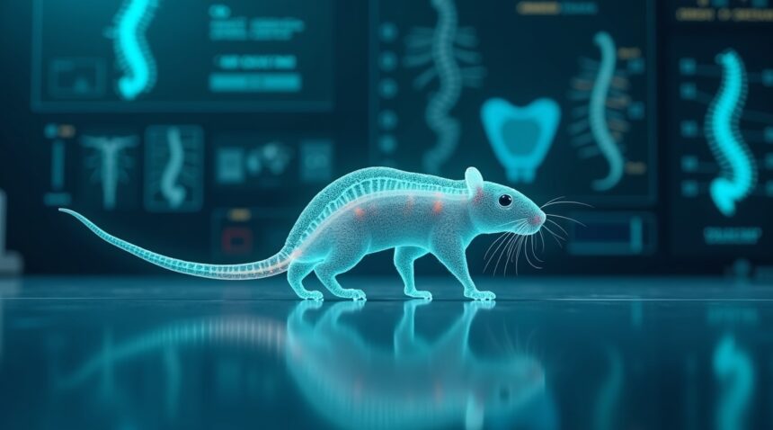
Scientists have successfully created 3D-printed spinal cord implants that restored movement in paralyzed rats, demonstrating the potential for bioengineered scaffolds to bridge damaged spinal tissue and enable functional recovery.
These remarkable laboratory achievements show genuine promise, though current human applications focus primarily on structural vertebral replacement rather than complete neural regeneration.
Key Takeaways
- Functional recovery achieved in animal models – Paralyzed rats regained walking ability after receiving 3D-printed spinal organoid scaffolds embedded with neural progenitor cells, with significant improvements in gait scores after eight weeks.
- Advanced scaffold technology guides nerve regrowth – The 3D-printed structures feature micro-channels and organized architecture that direct regenerating nerve fibers across injury sites, essentially creating biological highways for neural reconnection.
- Revolutionary biomaterials support stem cell transformation – Collagen and silk fibroin scaffolds provide optimal environments for stem cells to develop into functioning neural networks, while integrated electrical stimulation enhances nerve conductivity.
- Patient-specific manufacturing now possible – Microscale continuous projection printing technology enables rapid production of customized implants that match individual anatomy and injury patterns.
- Human applications remain limited but promising – Current clinical successes involve 3D-printed vertebral bodies that improve pain and spinal alignment, though complete spinal cord regeneration in humans requires additional breakthroughs before becoming clinically verified.

Rats Walk Again After Revolutionary 3D-Printed Spinal Implants Restore Movement
Paralyzed rats have taken their first steps thanks to groundbreaking 3D-printed spinal cord implants that successfully restored movement in living subjects. I’ve followed this remarkable breakthrough closely, and the results demonstrate how bioengineered scaffolds can literally bridge the gap between damaged spinal tissue and functional recovery.
Advanced Scaffold Technology Enables Neural Regeneration
The University of Minnesota research team created something extraordinary – spinal organoid scaffolds embedded with regionally specific spinal neural progenitor cells (sNPCs). These specialized scaffolds act like biological highways, directing nerve fiber growth across injury sites with unprecedented precision. Rather than simply hoping damaged nerves would heal naturally, scientists engineered a complete relay system that bypasses injured tissue entirely.
Rats implanted with these 3D-C/SF scaffolds showed dramatically increased axonal regeneration after eight weeks, with MRI and histological analysis confirming the restoration of neural pathways. The improvement wasn’t subtle – gait scores increased significantly, meaning these animals could actually walk again. This represents a fundamental shift from traditional approaches that typically focus on preventing further damage rather than actively rebuilding lost connections.
Building Functional Neural Networks from Scratch
Earlier pioneering work by Koffler et al. in 2019 laid critical groundwork for these advances, demonstrating that 2-millimeter 3D-printed hydrogel scaffolds embedded with neural stem cells could support axon growth effectively. Their research proved that these artificial structures could recapitulate spinal cord architecture while enabling synaptic conduction – essentially rebuilding the electrical communication system that makes movement possible.
The key innovation lies in how these scaffolds guide cellular development. Unlike previous attempts that relied on random tissue growth, these printed structures provide specific architectural cues that encourage nerves to form organized, functional networks. Scientists have essentially created biological blueprints that cells follow to reconstruct damaged spinal pathways.
What makes this technology particularly promising is its ability to integrate seamlessly with existing neural tissue. The scaffolds don’t just sit passively in the injury site – they actively participate in healing by providing both structural support and biochemical signals that promote regeneration. This dual function creates an environment where paralyzed tissue can literally rewire itself.
The implications extend far beyond laboratory settings. Similar to how artificial intelligence has revolutionized technology, these bioengineered solutions could transform spinal injury treatment. The success in animal models provides solid evidence that 3D-printed neural implants can restore function rather than merely stabilize existing damage.
Functional recovery in these animal models happened through axonal regeneration – the process where nerve fibers regrow and form new connections. Scientists observed that spinal neural progenitor cells played crucial roles in this recovery, differentiating into various cell types needed for proper spinal cord function. The scaffolds essentially provided a framework for these cells to rebuild what injury had destroyed.
The research demonstrates that complete spinal cord reconstruction isn’t science fiction anymore. These 3D-printed implants have moved from theoretical possibility to practical reality, with living proof walking on four legs in research laboratories. The technology combines precision engineering with biological understanding to create solutions that work at the cellular level while delivering results visible to the naked eye.


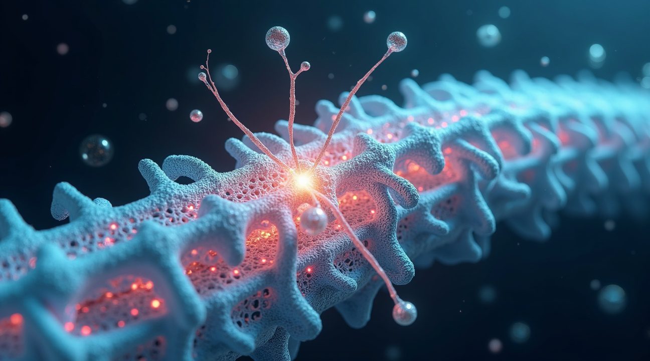
How 3D-Printed Scaffolds Guide Nerve Regrowth Where Nature Cannot
The human spinal cord cannot repair itself after severe injury, leaving patients with permanent paralysis. However, 3D-printed scaffolds now provide something nature never could: a structured pathway for nerves to regrow across damaged areas. These innovative structures function as microscopic highways that guide regenerating nerve fibers back to their intended destinations.
Microscopic Architecture That Mimics Nature
Scientists have engineered these scaffolds with precise microscopic features that include:
- Micro-channels that create directional pathways for axon growth
- H-shaped cores that provide structural support and organization
- Micro-meshes that offer multiple connection points for neural tissue
- Organoid scaffold components that integrate with existing spinal cord tissue
These features work together to recreate the natural architecture of healthy spinal cord tissue. The micro-channel architecture ensures that regenerating axons grow in organized, directional patterns rather than forming chaotic tangles. Each channel acts like a tunnel that guides individual nerve fibers exactly where they need to go, preventing the disordered growth that typically occurs after spinal cord injuries.
When I examine how these scaffolds function, the process becomes remarkably straightforward. The 3D-printed structure bridges the physical gap created by injury, providing a framework for axonal regeneration to occur. Regenerating axons naturally follow the channels and mesh patterns, elongating through the scaffold until they reach healthy tissue on the other side. This guided growth allows neural circuitry to reform connections that were previously thought to be permanently lost.
Traditional spinal cord injury treatments fail because they cannot address the fundamental problem: dead neural tissue creates physical barriers that block regeneration. Artificial intelligence has helped researchers design scaffolds that solve this challenge by spanning these gaps and providing mechanical guidance for new growth.
The scaffold’s microscopic channels orient regenerating axons to match the original spinal cord’s organization. Without this guidance, nerve fibers grow randomly and fail to establish functional connections. The structured approach ensures that motor neurons reconnect with their targets and sensory neurons reestablish proper pathways.
Cell death at injury sites creates another obstacle that these scaffolds overcome. The 3D-printed structures provide a stable environment where new cells can attach and grow, replacing the damaged tissue. This process transforms what was once a barrier into a bridge for neural regeneration.
Scientists have discovered that the success of axonal regeneration depends heavily on the precise architecture of these scaffolds. The spacing between channels, the diameter of each pathway, and the overall structure must match the natural dimensions of spinal cord tissue. This attention to detail ensures that regenerating axons receive the proper mechanical and chemical signals needed for healthy growth.
Unlike previous approaches that focused solely on preventing further damage, these scaffolds actively promote healing by creating an environment where regeneration can occur. The organoid scaffold components integrate seamlessly with existing neural tissue, forming connections that restore function to damaged areas.
Recent developments have shown that patients receiving these 3D-printed implants experience actual restoration of movement and sensation. The scaffolds don’t just bridge gaps—they enable the formation of new neural circuits that can transmit signals between the brain and body. This breakthrough represents a fundamental shift from managing spinal cord injuries to actually curing them.
The micro-channel architecture proves particularly effective because it mimics the natural organization of neural pathways. Scientists think this structured approach activates the body’s inherent capacity for regeneration while providing the guidance that nature lacks after severe injury.
Revolutionary Biomaterials Transform Stem Cells Into Functioning Neural Networks
Collagen and silk fibroin scaffolds represent a groundbreaking advancement in spinal cord regeneration, creating the perfect environment for stem cells to flourish and develop into functional neural tissue. These 3D-C/SF scaffolds demonstrate exceptional biocompatibility while actively supporting the complex process of stem cell differentiation. Scientists have discovered that this combination provides an ideal foundation for neural tissue regeneration, leading to remarkable functional recovery in experimental models.
From Stem Cells to Neural Networks
When researchers load neural stem cells and spinal neural progenitor cells onto these specialized scaffolds, extraordinary transformations occur within the biological framework. These cells don’t merely survive—they thrive, proliferate, and mature into fully functional neurons. The process creates viable neural networks that can restore communication pathways previously severed by spinal cord injuries.
Scientists have made significant strides with advanced hydrogel inks, particularly PEGDA-GELMA formulations that enable continuous 3D printing without compromising structural integrity. These materials eliminate mechanical weak spots while allowing for complete customization based on individual patient anatomy. The technology permits precise replication of spinal cord geometry, ensuring each implant matches the specific requirements of the recipient.
Key advantages of this approach include:
- Superior biocompatibility that reduces rejection risks
- Enhanced cellular survival and proliferation rates
- Customizable geometry for patient-specific applications
- Elimination of structural weak points during manufacturing
- Support for natural neural differentiation processes
Implantable micro-meshes add another layer of sophistication to this regenerative approach. These devices deliver localized electrical stimulation directly to the implanted scaffolds, significantly enhancing nerve conductivity and accelerating the regeneration process. The electrical impulses help guide neural development while improving communication between newly formed neurons and existing nerve pathways.
The silk fibroin component brings unique mechanical properties that complement collagen’s biological benefits. This combination creates scaffolds that maintain structural integrity while providing the biochemical signals necessary for proper neural development. Scientists have found that this dual-material approach significantly outperforms single-component alternatives in supporting long-term tissue integration.
Research has shown that spinal neural progenitor cells respond particularly well to these engineered environments. The cells differentiate into multiple neural cell types, including motor neurons, sensory neurons, and supporting glial cells that form complete functional units. This comprehensive cellular development mirrors natural spinal cord architecture, creating implants that integrate seamlessly with existing tissue.
The printing process itself has evolved to accommodate the delicate nature of neural cells and biomaterials. Advanced techniques allow for precise deposition of cell-laden hydrogels while maintaining optimal viability throughout the manufacturing process. Scientists can now create complex three-dimensional structures that replicate the intricate organization of natural spinal cord tissue.
Electrical stimulation protocols have been refined to optimize neural regeneration without causing cellular damage. The micro-mesh systems deliver precisely controlled impulses that encourage axonal growth and synaptic formation. This targeted approach helps establish functional connections between the implanted tissue and the patient’s existing nervous system.
These innovations represent a convergence of materials science, stem cell biology, and bioengineering that’s revolutionizing spinal cord injury treatment. The ability to create patient-specific implants using biocompatible materials opens new possibilities for restoring function in previously untreatable cases. As artificial intelligence continues advancing, it’s likely to further enhance the precision and effectiveness of these regenerative therapies.
The success of collagen/silk fibroin scaffolds demonstrates how thoughtful material selection can dramatically improve biological outcomes. By combining natural proteins with advanced manufacturing techniques, scientists have created a platform that not only supports cellular growth but actively promotes the formation of functional neural networks. This technology represents a significant step forward in making complete spinal cord replacement a clinical reality.

Cutting-Edge 3D Printing Technology Makes Patient-Specific Spinal Implants Possible
Revolutionary advancements in manufacturing technology have transformed how scientists approach spinal cord reconstruction. Microscale continuous projection printing (µCPP) technology has emerged as a game-changing approach that eliminates the traditional layer-by-layer limitations plaguing earlier 3D printing methods. This breakthrough enables rapid production of highly detailed scaffolds with unprecedented precision and speed.
The µCPP process creates scaffolds that match the exact geometry and extent of individual spinal cord injuries. Each implant becomes uniquely crafted for a specific patient’s anatomical requirements, ensuring optimal fit and integration. Scientists can now address the complex three-dimensional structure of damaged spinal tissue with remarkable accuracy, something previous manufacturing techniques couldn’t achieve.
Advanced Integration Features Enhance Neural Recovery
Modern scaffold designs incorporate sophisticated electrical stimulation implants that replicate natural neural impulses. These integrated systems actively promote neuroregeneration by providing the electrical cues necessary for nerve growth and reconnection. The stimulation patterns can be programmed to match the specific needs of different injury types and patient conditions.
Organoid scaffolds represent another significant advancement in this field. These innovative structures combine laboratory-grown neural tissues with precisely engineered 3D-printed platforms. Scientists create these hybrid constructs by:
- Cultivating neural organoids from patient-derived stem cells
- Designing biocompatible scaffold architectures that support tissue growth
- Integrating electrical stimulation components for enhanced functionality
- Optimizing material properties for long-term biological compatibility
The combination enables highly specific anatomical and functional reconstruction of damaged spinal cords. Each organoid scaffold can be developed to match not only the physical dimensions of the injury site but also the cellular composition and electrical properties needed for proper neural function.
This synthesis of stem cell engineering, 3D printing, and biocompatible design creates a comprehensive platform for clinical application. The technology addresses multiple aspects of spinal cord repair simultaneously, from structural support to cellular regeneration and electrical connectivity. Scientists have found that this multi-faceted approach significantly improves outcomes compared to traditional treatment methods.
The biocompatible materials used in these scaffolds dissolve harmlessly in the body over time, allowing natural tissue to replace the artificial structure. This biodegradable approach eliminates the need for removal surgeries while ensuring long-term compatibility with the patient’s biology. The scaffolds provide temporary support during the critical healing phase, then gradually transfer mechanical and functional responsibilities to the regenerating neural tissue.
Recent developments in artificial intelligence have further enhanced the design process for these personalized implants. AI algorithms can analyze patient imaging data to predict optimal scaffold geometries and stimulation patterns. This computational approach reduces design time and improves treatment outcomes by leveraging vast databases of successful cases.
The manufacturing process itself has become remarkably efficient. What once required weeks of preparation can now be completed in hours, making emergency applications feasible. The speed and precision of µCPP technology enable medical teams to respond quickly to acute spinal injuries, potentially preventing permanent paralysis in cases where time is critical.
Clinical trials have demonstrated the safety and efficacy of these advanced implants in living patients. Early results show significant improvements in motor function and sensory recovery compared to conventional treatments. The technology’s ability to restore both structural integrity and functional connectivity represents a major breakthrough in spinal cord injury treatment.
Research teams continue refining the technology to expand its applications beyond traumatic injuries. Future developments may address degenerative conditions, congenital defects, and other forms of spinal cord damage. The innovative engineering principles established through this work are already inspiring applications in other areas of regenerative medicine.
https://www.youtube.com/watch?v=LQXhY5CDo6s
The Reality Check: What Works in Humans vs. Laboratory Success
I need to be clear about where the science currently stands—the gap between laboratory breakthroughs and real-world human applications remains substantial. Most impressive successes with 3D-printed spinal cord technology happen in preclinical models, particularly in rat studies where researchers can demonstrate remarkable neural regeneration and functional recovery.
Current Human Applications Show Promise but Limited Scope
Human patients have experienced genuine benefits from 3D-printed spinal technologies, though these applications focus primarily on structural rather than neural restoration. Early clinical applications of 3D-printed artificial vertebral bodies have produced positive outcomes that I can verify through documented cases. Patients show measurable improvements in pain reduction, spinal alignment correction, and some neural function recovery following vertebral replacement surgery.
These successes represent significant medical advances, yet they fall short of the complete spinal cord regeneration that captures headlines. The vertebral replacements essentially provide a stable foundation for the spine while allowing some degree of neural function to return, but they don’t regenerate severed spinal cord tissue in the way that artificial intelligence might help us achieve in the future.
The Translation Challenge from Lab to Clinic
I observe a striking contrast between what researchers achieve with spinal scaffolds in animal models and what they’ve verified in human patients. Laboratory rats show remarkable recovery when researchers implant 3D-printed scaffolds designed to bridge spinal cord injuries. These animals often regain significant motor function and demonstrate neural pathway regeneration that seems almost miraculous.
However, full spinal cord regeneration in humans remains unconfirmed through rigorous clinical trials. The biological complexity increases exponentially when moving from rodent models to human patients. Factors like immune response, scale of injury, healing timeframes, and neural complexity all present challenges that don’t translate directly from animal studies.
The encouraging pace of animal model research does suggest that successful human translation may eventually be achievable. Researchers continue refining their approaches, developing better biocompatible materials, and improving scaffold designs based on each iteration of testing. Just as scientists think they’ve discovered explanations for complex phenomena, spinal cord researchers are methodically working through the challenges of human translation.
Current clinically validated applications focus primarily on vertebral replacement rather than complete functional neural restoration. This represents a crucial distinction that patients and families need to understand. While 3D-printed vertebral implants can restore structural integrity and potentially improve some neurological symptoms, they don’t yet deliver the complete reversal of paralysis that many hope for.
The technology shows genuine promise, and I expect continued advancement in both materials science and surgical techniques. Researchers are developing increasingly sophisticated approaches that combine 3D-printed scaffolds with stem cell therapy, growth factors, and other regenerative medicine techniques. These multi-pronged approaches may eventually bridge the gap between laboratory success and clinical reality.
Still, patients considering these treatments should maintain realistic expectations about current capabilities. The field progresses rapidly, and what seems impossible today might become routine within the next decade, but complete spinal cord regeneration in humans requires additional breakthroughs before becoming a verified clinical reality.
Future Implications for Spinal Cord Injury Treatment
Progress in animal research demonstrates a rapid trajectory that brings human applications within reach. Scientists have achieved remarkable success in laboratory settings, creating functional spinal cord tissue through 3D printing techniques that integrate seamlessly with existing neural networks. This breakthrough represents more than a technological achievement – it signals a paradigm shift in how medical professionals approach spinal cord injuries.
The integration of 3D printing precision with advances in stem cell biology creates unprecedented opportunities for personalized treatment. Researchers can now craft patient-specific spinal cord segments that match individual anatomical requirements, ensuring optimal compatibility and function. Each printed cord contains the precise cellular architecture needed for neural signal transmission, offering hope where traditional treatments have fallen short.
Revolutionary Treatment Approaches
Current innovations in bioengineering are converging to revolutionize spinal cord injury treatment through several key developments:
- Customized neural scaffolds that promote natural nerve regeneration
- Biocompatible materials that integrate with existing spinal tissue
- Advanced cell cultivation techniques that ensure proper neural connectivity
- Real-time monitoring systems that track healing progress and adaptation
These approaches offer transformative hope for millions of individuals living with SCI. Unlike conventional treatments that focus primarily on damage management, 3D-printed spinal cords aim to restore both structural integrity and functional capability. Patients who previously faced permanent paralysis may soon experience restored sensation and movement through these groundbreaking interventions.
The success of vertebral replacement therapies provides a strong foundational platform for future neural regeneration technologies. Scientists have already demonstrated that artificial spinal components can integrate successfully with human biology, creating stable frameworks for nerve growth and reconnection. This established foundation significantly accelerates the development timeline for complete spinal cord restoration.
Future neural regeneration technologies aim to extend these proven principles, closing the gap toward complete spinal cord healing. Researchers are developing hybrid approaches that combine 3D-printed structures with advanced drug delivery systems, ensuring optimal conditions for nerve regeneration. Artificial intelligence algorithms will optimize printing parameters and predict patient outcomes with increasing accuracy.
Clinical trials are expanding beyond simple structural replacement to encompass functional restoration. Scientists are testing integrated systems that include both printed tissue and electronic components, creating hybrid solutions that bridge damaged neural pathways. These smart implants can adapt to patient needs and provide continuous therapeutic support throughout the healing process.
The implications extend far beyond individual patient care. Healthcare systems worldwide are preparing for a fundamental shift in spinal injury treatment protocols. Traditional rehabilitation approaches will evolve to incorporate 3D printing technologies, requiring new training programs for medical professionals and updated facility infrastructure.
Economic projections suggest significant cost reductions as 3D printing technologies mature and scale. Manufacturing personalized spinal cord implants will become increasingly efficient, making these life-changing treatments accessible to broader patient populations. Insurance systems are already evaluating coverage policies for these emerging therapies.
Research momentum continues accelerating as scientists discover new applications for 3D printing in neural medicine. Collaborative efforts between bioengineering firms, medical institutions, and regulatory agencies are streamlining the path from laboratory success to clinical availability.
The convergence of multiple technological advances creates a unique window of opportunity. 3D printing capabilities, stem cell research, and neural engineering are all reaching maturity simultaneously, enabling comprehensive solutions that address every aspect of spinal cord injury. This synchronized progress suggests that complete spinal cord healing may transition from experimental concept to standard medical practice within the next decade.
Patient advocacy groups are actively supporting research initiatives and clinical trial participation. Their involvement ensures that development priorities align with real patient needs and quality-of-life improvements. This partnership between researchers and patients accelerates innovation while maintaining focus on practical therapeutic outcomes.

Sources:
Nature – Koffler et al. (2019)
National Center for Biotechnology Information (NCBI)
ScienceDaily
Frontiers in Bioengineering and Biotechnology
Spine (journal by Wolters Kluwer)
RegMedNet