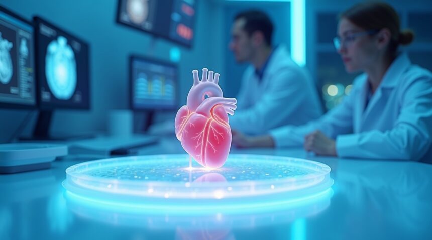
Israeli researchers at Tel Aviv University achieved a groundbreaking milestone in regenerative medicine by successfully 3D printing the world’s first living, beating human heart using only a patient’s own biological materials.
Key Takeaways
- Researchers successfully 3D printed a complete living heart with functional chambers, blood vessels, and beating cardiac cells using only the patient’s own fat tissue.
- The personalized approach eliminates immune rejection risks since the printed organ originates entirely from the patient’s biological material.
- The four-step process transforms fat cells into heart tissue through cellular reprogramming, bio-ink development, and precise 3D printing guided by medical imaging.
- Current applications will focus on cardiac patches for heart repair within five years, with complete organ replacements potentially available within the next decade.
- This breakthrough addresses the critical organ shortage crisis, where only 4,000 heart transplants occur globally each year while thousands of patients die waiting for donors.
The Science Behind the Breakthrough
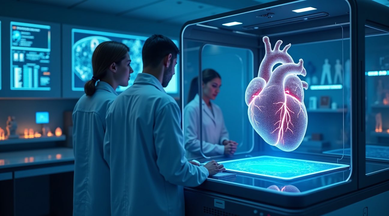
The research team extracted fat cells from patients and performed cellular reprogramming to convert them into cardiac muscle cells and blood vessel cells. Scientists then combined these reprogrammed cells with specialized bio-inks to create a printable material. The team used CT scans and MRI imaging to map the patient’s heart structure, providing precise blueprints for the 3D printing process.
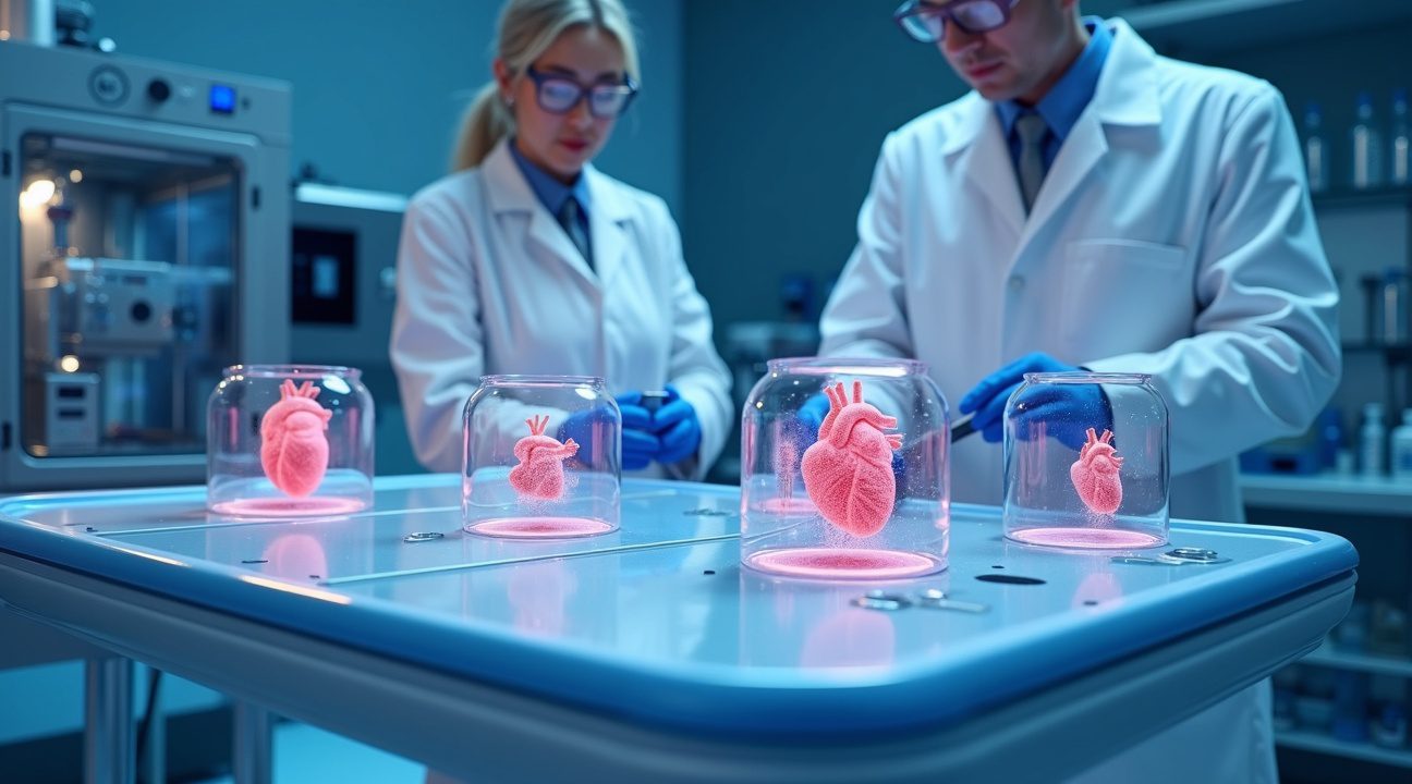
The printed heart measures approximately 2.5 centimeters and weighs about 10–15 grams. While current technology produces hearts suitable for small animals, researchers expect rapid scaling to produce human-sized organs. The organ demonstrates actual cardiac function, with cells contracting in synchronized patterns similar to natural heart rhythms.
Advantages Over Traditional Transplants
This technology offers several significant advantages over traditional heart transplants. Patients won’t need lifelong immunosuppressive medications, which often cause serious side effects. The approach eliminates waiting lists for donor organs, allowing immediate treatment for acute cardiac conditions.
Short-Term Applications
Clinical trials for cardiac patches are expected to begin within five years. These patches can repair damaged heart tissue resulting from heart attacks or congenital defects.
Long-Term Vision
Complete heart replacement procedures require further development, but show promise for becoming viable within the next decade. This advancement may redefine long-term outcomes for patients with end-stage heart failure.
Addressing a Global Health Crisis
Heart disease remains the leading cause of death globally. Current treatments, such as artificial hearts, offer only temporary solutions and lack the biological integration necessary for long-term success. This innovation addresses a massive medical need by offering a fully functional, patient-specific solution.
Challenges and Future Developments
Manufacturing costs remain high for individual organs, but researchers anticipate significant cost reductions as the technology matures. Currently, the process takes several weeks from cell extraction to the completed organ. However, increased automation will likely shorten production times significantly.
Future efforts include creating larger organs suitable for adult patients. Researchers are also refining bio-ink formulations to improve cell survival and organ durability. Integration with existing surgical practices will require further research to ensure successful implantation procedures.
A Transformational Shift in Cardiac Medicine
This achievement represents a fundamental transformation in the field of cardiac care: a shift from organ transplantation to organ manufacturing. The technology developed by the Tel Aviv University team has the potential to radically improve outcomes for millions of individuals suffering from cardiovascular disease, and offers new hope for conditions once considered untreatable.

Tel Aviv University Scientists Create World’s First Living, Beating 3D Printed Heart
In April 2019, researchers at Tel Aviv University achieved something that seemed impossible just decades ago. Prof. Tal Dvir of Tel Aviv University’s School of Molecular Cell Biology and Biotechnology led a groundbreaking team that successfully 3D printed the world’s first living, beating human heart using entirely biological materials from a single patient.
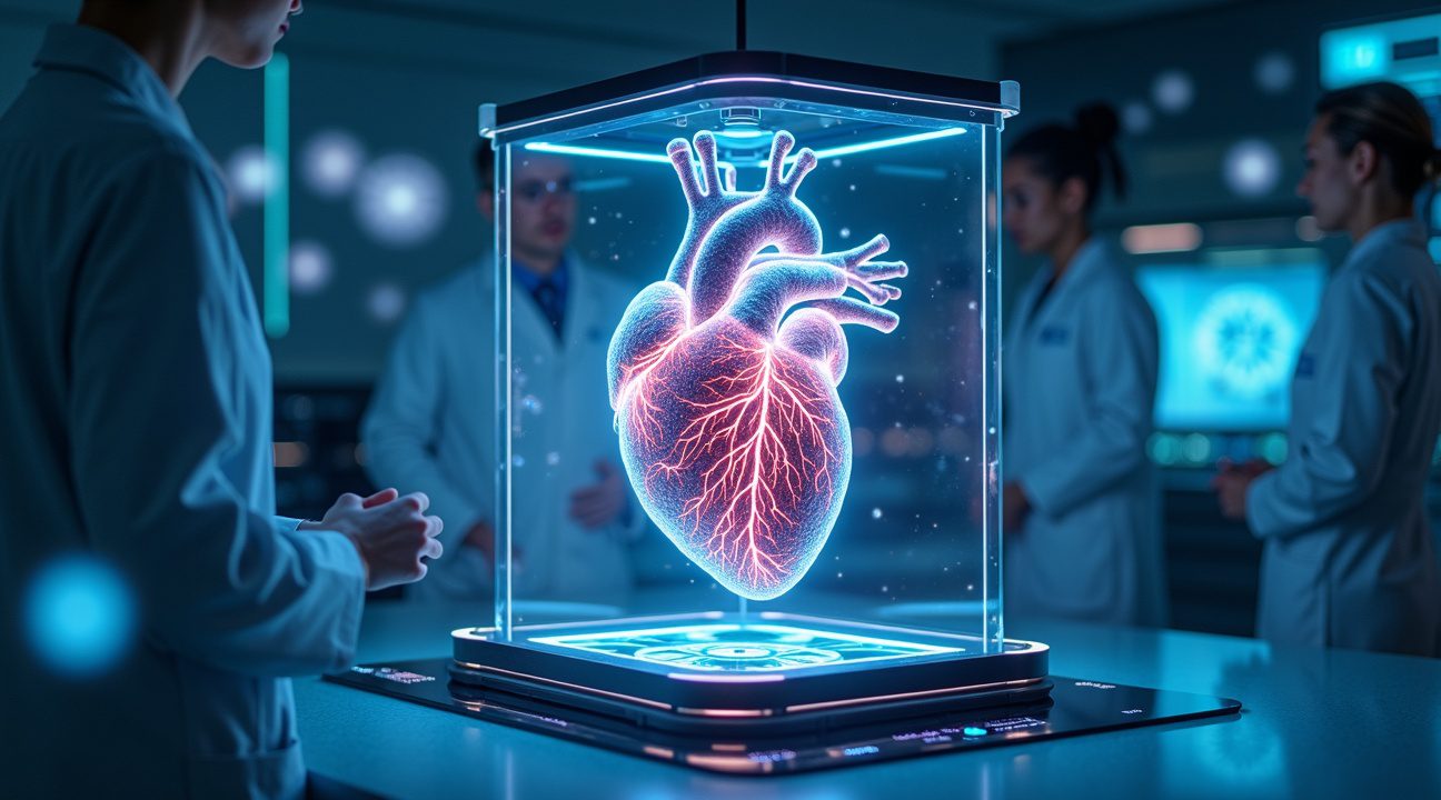
This achievement represents a massive leap forward in regenerative medicine and organ transplantation. Unlike previous attempts that could only produce simple tissues or non-living structures, I can confirm this heart contained all the essential components of a functioning organ. The printed heart included living heart cells, complete blood vessel networks, ventricles, and chambers that could actually contract and beat.
Technical Specifications and Process
The printed organ measured approximately 2.5 cm (1 inch) in size, making it about the size of a rabbit’s heart. While this might seem small compared to an adult human heart—roughly 100 times smaller—it demonstrated that the technology could successfully create complex cardiac tissue with proper cellular organization and vascularization.
The entire printing process required just three hours to complete, which is remarkably fast considering the complexity of the organ being created. The researchers used the patient’s own cells and biological materials, which could potentially eliminate the risk of immune rejection that plagues traditional organ transplants. This personalized approach means each printed heart would be perfectly matched to the recipient’s biological profile.
What makes this achievement particularly significant is the successful integration of multiple tissue types and structures. Previous 3D bioprinting efforts struggled to create organs with proper blood vessel networks, but this heart included functional vascularization throughout the tissue. The chambers and ventricles were properly formed, and the cardiac cells demonstrated the ability to contract rhythmically, creating the characteristic beating motion of a living heart.
This breakthrough opens new possibilities for patients suffering from heart disease, which remains one of the leading causes of death worldwide. While the current size limitation means human applications are still years away, the proof of concept shows that scientific breakthroughs can rapidly transform from theoretical concepts into practical realities. The research team’s success in creating a fully vascularized organ addresses one of the biggest challenges in tissue engineering and brings us significantly closer to solving the global organ shortage crisis.

Revolutionary Bio-Ink Technology Uses Patient’s Own Cells to Prevent Rejection
I find this breakthrough particularly fascinating because it addresses one of the most persistent challenges in organ transplantation. Previous 3D printed organ models fell short of clinical viability, producing basic tissues that lacked blood vessels or living, beating cells. Scientists struggled to create functional organs that could actually support life once implanted.
This printed heart represents a monumental leap forward as the first fully vascularized and cellular 3D printed organ. The researchers developed patient-specific bio-inks composed entirely of the individual’s own tissue, including essential sugars and proteins. This personalized approach ensures complete immunological compatibility between the printed organ and the recipient’s body.
Transforming Fat Cells Into Heart Tissue
The process begins with a remarkably simple procedure. I learned that researchers need only a small fat tissue biopsy from the patient to create this life-saving organ. They then convert these fat cells into cardiac and endothelial cells using advanced stem cell technologies. This transformation creates all the necessary cellular components for a functioning heart.
The printed heart achieves both anatomical and cellular matching to the patient’s real heart. This precision ensures that the organ integrates seamlessly with the recipient’s cardiovascular system. The bio-ink contains the exact cellular makeup and structural proteins needed to support proper heart function.
Eliminating Immunosuppressive Drug Dependency
Current heart transplant recipients face a lifetime of immunosuppressive medications to prevent organ rejection. These drugs carry significant side effects and leave patients vulnerable to infections and certain cancers. The use of patient-derived tissue in this 3D printing process could eliminate this burden entirely.
Since the printed heart originates from the patient’s own cells, the immune system recognizes it as native tissue rather than foreign material. This recognition prevents the typical rejection response that necessitates ongoing immunosuppressive therapy. Patients could potentially receive their new heart without the need for any anti-rejection medications.
The innovation extends beyond just preventing rejection. The patient-specific approach means each heart is perfectly sized and shaped for its intended recipient. Traditional transplants often involve size mismatches between donor and recipient, leading to surgical complications and suboptimal outcomes.
This technology represents a significant advancement over previous attempts at scientific breakthroughs in organ engineering. The researchers overcame fundamental limitations that had prevented earlier 3D printed organs from achieving clinical relevance.
The fully vascularized nature of this printed heart means it contains the complete network of blood vessels necessary for proper circulation. Previous attempts created solid tissue masses that couldn’t support adequate blood flow. This new approach ensures that every part of the printed heart receives the oxygen and nutrients it needs to function properly.
The cellular components beat spontaneously, demonstrating that the printed tissue maintains the electrical properties essential for heart function. This beating action proves that the printed organ possesses the fundamental characteristics needed to pump blood throughout the body.
Most importantly, the entire process relies exclusively on the patient’s biological material. No synthetic scaffolds or foreign substances are required, further reducing the risk of adverse reactions. This approach represents a true autologous transplant, where the patient essentially receives an upgraded version of their own tissue.
The potential impact of this technology extends far beyond individual patients. Healthcare systems worldwide could benefit from reduced costs associated with long-term immunosuppressive therapy and its related complications. The technology could also address the critical shortage of donor hearts that currently limits transplant options for thousands of patients.

Four-Step Process Transforms Fat Tissue into Beating Heart Chambers
The revolutionary technique developed by Israeli researchers follows a precise four-step methodology that transforms a patient’s own fat tissue into a fully functional cardiac organ. This groundbreaking approach eliminates the risk of immune rejection while creating hearts that match each patient’s unique anatomy.
Tissue Harvesting and Cellular Separation
The process begins with a simple biopsy procedure where doctors extract a small sample of fatty tissue from the patient. I find this initial step remarkable because it uses readily available adipose tissue rather than requiring more invasive procedures. The harvested fat undergoes careful separation to isolate two critical components: the cellular material and the extracellular matrix. This separation forms the foundation for everything that follows, as each component serves a distinct purpose in the heart creation process.
Cellular Reprogramming and Bio-ink Development
The separated cells then undergo an extraordinary transformation through reprogramming into pluripotent stem cells. These versatile cells can develop into any type of tissue, allowing researchers to differentiate them into two essential heart cell types. The process creates cardiomyocytes, which form the heart muscle and generate the beating motion, alongside endothelial cells that line the blood vessels. Meanwhile, the extracellular matrix receives processing treatment to become a specialized hydrogel bio-ink.
The combination of reprogrammed cells with the bio-ink creates a printable material that maintains cellular viability while providing structural support. This innovative bio-ink formulation represents a significant advancement over previous attempts that relied on synthetic materials or foreign biological components.
3D Printing with Anatomical Precision
The final stages leverage advanced medical imaging technology to ensure anatomical accuracy. CT or MRI scans from the patient provide detailed blueprints that guide the 3D printer’s construction process. The printer follows these precise specifications to build ventricles, chambers, and intricate vascular networks that mirror the patient’s original cardiac architecture.
This personalized approach addresses one of the most challenging aspects of heart transplantation: compatibility. Rather than hoping for a donor match, the process creates organs specifically designed for each individual patient. The resulting hearts contain living, beating chambers that demonstrate the same contractile properties as natural cardiac tissue.
The technique represents a convergence of multiple scientific disciplines, from cellular biology to advanced manufacturing. Each step builds upon the previous one, creating a seamless progression from simple fat tissue to a complex, functioning organ. The success of this method opens new possibilities for treating heart disease while potentially eliminating lengthy transplant waiting lists. Similar breakthroughs in biotechnology continue to push boundaries, much like recent developments in particle research that expand our understanding of biological systems.
Critical Medical Need Drives Urgency for Organ Printing Breakthrough
Heart disease continues to claim more lives than any other medical condition globally, creating an urgent demand for revolutionary treatment solutions. I’ve observed how current therapeutic options remain severely limited for patients facing end-stage heart failure, with heart transplantation standing as the only viable treatment for these critical cases.
The Devastating Organ Shortage Crisis
The statistics paint a grim picture of the current organ shortage crisis. Surgeons perform only around 4,000 heart transplants globally each year, leaving countless patients without hope. The United States alone witnesses approximately 20 people dying daily while waiting for a suitable donor heart. These numbers highlight why researchers have turned to innovative scientific breakthroughs in 3D bioprinting technology.
This scarcity stems from multiple factors, including:
- The complex matching requirements between donors and recipients
- Geographical constraints
- The limited time window for organ viability after harvest
Traditional organ procurement relies on tragic circumstances that provide healthy organs, creating an inherently unpredictable and insufficient supply chain.
Bridging Technology: Cardiac Patches as First Steps
Early applications of this groundbreaking technology will focus on producing cardiac patches rather than complete organ replacements. These patches can repair damaged areas of the heart, offering immediate therapeutic benefits while researchers continue developing full-scale organ printing capabilities. This approach represents a practical stepping stone that could help countless patients before complete heart printing becomes feasible.
The transition from patches to full organs addresses one of the most significant current challenges in bioprinting: teaching these 3D printed hearts to contract and pump blood effectively. Scientists must ensure these printed organs function reliably over extended periods in animal studies before advancing to human trials. This process requires extensive testing to verify that the printed cardiac tissue:
- Maintains proper electrical conductivity
- Responds appropriately to physiological signals
- Demonstrates long-term structural integrity
I’ve noted that this technology’s development timeline reflects both the complexity of creating functional cardiac tissue and the rigorous safety standards required for human implantation. Each advancement brings researchers closer to eliminating organ waiting lists entirely, potentially transforming cardiovascular medicine and saving millions of lives annually.
Timeline and Future Applications Within Next Decade
Prof. Dvir’s predictions paint an optimistic picture for the future of regenerative medicine. According to his projections, personalized 3D-printed organs may become available in leading hospitals within the next 10 years. This timeline reflects the rapid advancement in bioprinting technology that has accelerated dramatically since the initial breakthrough.
The development path shows a clear progression in complexity. Simpler tissues and organs are expected to reach clinical application much earlier than complex structures like the heart. Skin patches, cartilage replacements, and basic tissue grafts will likely lead the charge in the coming years. More intricate organs requiring sophisticated vascular networks and multiple cell types will follow as the technology matures and regulatory frameworks adapt to these innovations.
Revolutionary Advancement Over Previous Methods
Previous attempts at bio-printing faced significant limitations that the Tel Aviv University team successfully overcame. Earlier research efforts were restricted to creating simple, non-living tissue structures or basic scaffolds without vascular or cellular functionality. These primitive approaches posed substantial rejection risks because they couldn’t integrate properly with the patient’s existing biological systems.
The breakthrough achieved by Israeli researchers represents a fundamental shift in approach. Tel Aviv University’s model produces a fully cellular, vascularized, and beating 3D printed heart that’s carefully tailored to the specific patient’s anatomy. This customization dramatically reduces immunological rejection risks since the organ originates from the patient’s own biological material. The heart contains all essential components: living cells, functional blood vessels, and properly formed chambers that contract rhythmically.
This technological leap addresses the most pressing challenges in organ transplantation:
- Current transplant procedures require lifelong immunosuppressive drug use.
- Traditional organs carry high rejection risks due to immune incompatibility.
- Patient-specific organs eliminate these challenges by reducing or removing immune response.
The implications extend far beyond cardiac applications. Similar techniques could revolutionize treatment for liver disease, kidney failure, and numerous other conditions requiring organ replacement. Scientists are already exploring applications for scientific breakthroughs in related fields that could accelerate development timelines.
Early clinical trials will likely focus on smaller, less complex applications before progressing to full heart replacements. Cardiac patches for repairing damaged heart tissue could become available within five years, offering hope to millions of patients with heart disease. These patches would serve as stepping stones, allowing researchers to refine techniques and gather safety data before attempting complete organ replacement.
The manufacturing process itself continues to evolve rapidly:
- Current production times are lengthy.
- Automation and improved printing technologies are expected to shorten printing duration.
- Costs, currently high, should decrease with scale and standardization.
Regulatory approval represents another crucial milestone on the path to clinical availability. Medical authorities worldwide are developing new frameworks to evaluate these unprecedented treatments. The complexity of assessing a living, patient-specific organ requires entirely new testing protocols and safety standards.
International collaboration accelerates progress as research teams share findings and techniques. The Israeli breakthrough has sparked increased investment in bioprinting research globally, with major medical centers establishing dedicated programs. This collaborative environment suggests that Prof. Dvir’s 10-year timeline could prove conservative if current momentum continues.
The technology’s potential to reshape regenerative medicine extends beyond organ replacement. Applications in drug testing, disease modeling, and personalized medicine research could transform how medical professionals approach treatment development. Each printed organ could serve as a unique testing platform for therapies specific to individual patients.
Patient-specific organ creation represents just the beginning of what’s possible. Future developments may enable on-demand organ printing in hospital settings, eliminating wait times entirely. This capability could transform emergency medicine, offering immediate solutions for trauma patients and those experiencing sudden organ failure.
https://www.youtube.com/watch?v=QOZSU2kDKsQ
Sources:
The Times of Israel, “Israeli scientists unveil world’s first 3D-printed heart with human tissue”
Tel Aviv University, “TAU scientists print first ever 3D heart using patient’s own cells”
CBS News, “3D-printed heart a potential breakthrough in making human organs”
Xinhuanet, “Feature: Israeli scientists use 3D printing to create world’s 1st model heart”
i24NEWS, “Israeli Scientists Unveil ‘first’ 3D Print Of Heart With Human Tissue …”
YouTube, “Israeli researchers print 3D heart using patient’s own cells”
YouTube, “Israeli Lab Creates 3D-Printed Hearts with Biotic Tissue”