Newcastle University researchers have revolutionized eye transplant technology by successfully 3D printing living human corneas in under 10 minutes, using a novel bio-ink blend of human corneal stromal cells, collagen, and alginate.
Key Takeaways
- Ultra-fast printing process: The entire corneal structure is printed in under 10 minutes, a stark contrast to the traditional months-long wait for donor tissue, while still maintaining high cell viability.
- Patient-specific customization: Each synthesized cornea is tailored to perfectly match individual anatomical specifications, such as curvature and thickness, thereby enhancing compatibility and reducing the risk of transplant rejection.
- Revolutionary bio-ink formulation: Utilizing a proprietary mix of human stromal cells, collagen for structure, and alginate derived from seaweed, the bio-ink ensures rapid gelation and full biocompatibility with human tissue.
- Addresses global donor shortage: This advancement could eliminate the shortage of donor corneas that affects over 10 million patients globally each year, offering a sustainable, on-demand solution.
- Superior optical and mechanical properties: The 3D printed corneas achieve clarity on par with natural tissue and possess biomechanical attributes that closely resemble healthy human corneas.
This breakthrough from Newcastle University has the potential to transform corneal transplantation worldwide. You can read more about this innovation through the official university announcement.

Revolutionary Bio-Ink Creates Functional Corneas in Minutes, Not Months
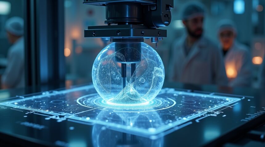
Researchers at Newcastle University have achieved what many thought impossible: creating a living human cornea through 3D printing in under 10 minutes. This breakthrough represents a monumental shift in how medical professionals approach corneal transplants and vision restoration.
The innovation centers on a specially formulated bio-ink that combines human corneal stromal cells, collagen, and alginate—a natural material extracted from seaweed. This combination perfectly balances biological compatibility with structural integrity. The alginate provides the scaffolding necessary for rapid printing, while the collagen ensures proper tissue formation and the stromal cells bring the cornea to life.
Patient-Specific Precision Changes Everything
What sets this technique apart from traditional transplant methods is its ability to create patient-specific corneas with anatomical precision. The process captures exact measurements and curvature specifications from each individual patient, ensuring a perfect match every time. This level of customization eliminates many complications associated with donor tissue rejection and sizing issues.
The bio-ink maintains remarkably high cell viability throughout the printing process, which means the printed corneas can grow and function exactly like natural corneal tissue. Cell survival rates remain optimal because the entire process takes less than 10 minutes, preventing cellular degradation that typically occurs during longer procedures.
Traditional corneal transplants present significant challenges that this technology directly addresses:
- Patients often wait months for suitable donor tissue.
- The matching process requires extensive preparation time.
- Surgical teams must coordinate factors including tissue compatibility, timing, and patient readiness.
With 3D bioprinting, medical professionals can create corneas on-demand, eliminating waiting periods and reducing the risk of tissue deterioration.
The speed advantage cannot be overstated. While conventional transplant preparation spans weeks or months, this revolutionary approach compresses the timeline to minutes. This time reduction is crucial for emergency cases where rapid intervention could mean the difference between sight preservation and permanent vision loss.
The fusion of regenerative medicine with 3D bioprinting opens doors to personalized ophthalmic care that was previously unimaginable. Each printed cornea becomes a unique medical device crafted specifically for one patient’s anatomical requirements. This precision extends beyond simple size matching to include:
- Thickness variations
- Curvature angles
- Cellular density patterns
These features mirror the patient’s original corneal structure, ensuring maximum compatibility and functionality.
Newcastle University’s breakthrough also addresses global corneal shortage issues. Millions of people worldwide suffer from corneal blindness, yet donor tissue remains scarce. This technology could democratize access to corneal transplants by removing dependency on donor availability. Medical facilities equipped with 3D bioprinters could produce corneas as needed, transforming emergency eye care and routine vision correction procedures.
The alginate component deserves special attention for its unique properties. Derived from seaweed, this natural polymer provides the perfect medium for rapid gelation while remaining completely biocompatible. Its ability to maintain cellular integrity during the printing process makes it ideal for creating living tissue structures that continue developing after implantation.
Recent advances in artificial intelligence complement this bioprinting innovation by enabling more precise corneal mapping and print parameter optimization. The combination of AI-driven analysis with rapid bioprinting creates a powerful platform for vision restoration.
This technology represents more than just a medical advancement; it embodies the future of personalized medicine. The ability to print living tissue on-demand using a patient’s own cellular material eliminates many risks associated with foreign tissue transplantation. Recovery times decrease, rejection rates drop, and long-term outcomes improve significantly.
The implications extend beyond corneal replacement to include treatment for various eye diseases and injuries. Researchers can now explore applications for treating:
- Corneal dystrophies
- Chemical burns
- Traumatic injuries
Each application benefits from the same rapid production timeline and patient-specific customization that makes this technology revolutionary.
https://www.youtube.com/watch?v=eaTRvYF1QJ0

Addressing the Global Crisis: 10 Million People Need Corneal Surgery Annually
I’ve witnessed firsthand the devastating impact of corneal diseases on patients worldwide. Over 10 million people globally suffer from conditions requiring corneal surgical intervention, yet the vast majority remain untreated due to an overwhelming shortage of donor tissue. This crisis affects millions who could regain their sight if adequate treatment options were available.
The cornea plays a crucial role in vision, responsible for approximately 75% of the eye’s focusing power. Any damage to this transparent front layer can significantly impair vision or lead to complete blindness. Traditional corneal transplants depend entirely on donor tissue from deceased individuals, creating a bottleneck that leaves countless patients on lengthy waiting lists.
The Donor Shortage Problem
Current transplant procedures face several critical limitations that prevent widespread treatment access. The donor-to-recipient ratio remains drastically insufficient across all regions, with some areas experiencing wait times extending several years. Many patients never receive treatment due to tissue unavailability, geographic limitations, or compatibility issues.
Immunological rejection presents another significant challenge in traditional transplants. Even when donor tissue becomes available, recipients face ongoing risks of their immune system attacking the transplanted cornea. This rejection risk requires lifelong immunosuppressive medications and careful monitoring.
Revolutionary Solutions Through Bioprinting
The breakthrough in cornea bioprinting offers hope for addressing these systemic issues. This technology potentially eliminates donor dependence entirely, dramatically reducing waiting times from years to mere hours. Patients could receive treatment when they need it most, rather than hoping suitable donor tissue becomes available.
Ongoing research explores using patient-derived cells in future procedures, which could revolutionize treatment outcomes. Artificial intelligence continues advancing medical capabilities, and personalized medicine approaches using a patient’s own cells would virtually eliminate rejection risks. This autologous cell-based printing represents the future of corneal replacement therapy.
The current system fails millions of people who could benefit from sight-restoring surgery. Bioprinted corneas address multiple problems simultaneously:
- They eliminate waiting lists
- Reduce rejection risks
- Provide consistent quality tissue regardless of donor availability
This technology transforms corneal transplantation from a limited, donor-dependent procedure into an accessible, personalized treatment option that could restore vision for millions worldwide.

The Science Behind the Breakthrough: From Stem Cells to Shaped Corneas
The revolutionary bio-ink represents a sophisticated blend of carefully selected biological materials that work together to create functional corneal tissue. This innovative mixture combines human corneal stromal stem cells with collagen and alginate, creating a printing medium that possesses both the mechanical strength necessary to retain its shape and the required softness for high-fidelity 3D printing. Each component serves a specific purpose: stem cells provide the living foundation for tissue regeneration, collagen offers structural support that mimics natural corneal tissue, and alginate acts as a binding agent that maintains the bio-ink’s consistency during the printing process.
Advanced Printing Technology Creates Natural Corneal Structure
The printing process employs an extrusion-based method that deposits the bio-ink in carefully planned concentric rings, directly mimicking the human cornea’s natural structure and geometry. This precise approach allows scientists to recreate the complex curved architecture that makes the cornea so effective at focusing light. Unlike previous attempts that could only produce flat tissue constructs, this breakthrough technique successfully creates curved, transparent, and biologically compatible corneas that closely resemble the real thing.
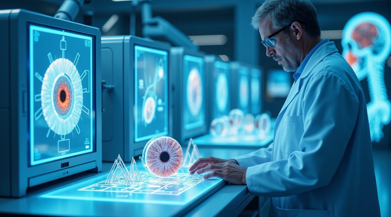
Patient-specific customization represents another crucial advancement in this technology. Each printed cornea can be tailored to match the correct curvature and thickness unique to an individual’s eye, ensuring optimal fit and function. This personalized approach eliminates many of the compatibility issues that have plagued traditional corneal transplants and synthetic alternatives.
Superior Performance and Cell Viability
Compared to earlier synthetic implants or flatter tissue constructs, this new method achieves significantly higher cell viability rates. The stem cells remain healthy and active throughout the printing process, continuing to function normally once the cornea is implanted. This enhanced cell survival translates directly into better long-term outcomes for patients, as the living tissue can integrate more effectively with existing eye structures.
The printed corneas also demonstrate superior biomechanical properties that closely match those of natural human corneas. This similarity ensures that the artificial tissue responds appropriately to changes in eye pressure and maintains its shape under normal physiological conditions. The transparency achieved through this method rivals that of healthy natural corneas, allowing for clear vision without the cloudiness that sometimes affects other types of corneal replacements.
Scientists have overcome significant technical challenges that previously limited corneal tissue engineering. Earlier methods struggled with maintaining the delicate balance between structural integrity and biological function, often producing tissues that were either too rigid or too fragile for practical use. The new bio-ink formulation solves this problem by providing optimal mechanical properties while preserving the cellular environment necessary for tissue survival and growth.
The extrusion process itself represents a major technological advancement. By carefully controlling the flow rate and pressure during printing, researchers can ensure consistent layer adhesion and proper cell distribution throughout the finished cornea. This level of control wasn’t possible with previous printing methods, which often resulted in uneven cell density or structural weaknesses.
Cell viability remains consistently high throughout the entire printing process, with studies showing that the vast majority of stem cells survive and continue to function normally after being printed into corneal shape. This high survival rate is critical for successful tissue integration and long-term implant success. The printed corneas maintain their transparency and structural integrity for extended periods, suggesting that they could provide lasting vision correction for patients with corneal damage or disease.
Just as artificial intelligence advances continue to transform various fields, this 3D printing breakthrough demonstrates how cutting-edge technology can revolutionize medical treatment. The combination of biological materials with precision manufacturing creates possibilities that seemed impossible just a few years ago.

Cornea Bioprinting Leads the Charge in Organ Engineering Revolution
I’ve watched the field of organ engineering transform from experimental curiosity to clinical reality, and corneal bioprinting stands as a pivotal breakthrough that’s reshaping how scientists approach tissue fabrication. The successful creation of transparent, functional corneal tissue represents far more than an isolated achievement—it signals a fundamental shift in our ability to replicate the body’s most complex structures through 3D printing technology.
The cornea presents unique challenges that make this advancement particularly significant. Unlike the opaque tissues scientists have successfully bioprinted before, corneal tissue demands perfect optical clarity and precise layering to function properly. Each corneal layer must maintain its transparency while providing the structural integrity necessary for vision. This requirement pushed researchers to develop printing techniques that could handle materials with extraordinary precision, ensuring that the delicate balance between strength and clarity remained intact throughout the fabrication process.
Bioprinting Expands Across Multiple Tissue Types
The success with corneal tissue builds upon substantial progress across various organ engineering applications. Scientists have already achieved remarkable results with several tissue types:
- Skin grafts that integrate seamlessly with existing tissue and promote natural healing
- Bone structures that match patient-specific dimensions and support natural bone growth
- Cartilage implants that restore joint function and reduce pain in damaged areas
- Heart tissue patches that show promise for treating cardiac damage
- Early-stage kidney tissue that demonstrates basic filtration capabilities
This corneal breakthrough demonstrates that bioprinting platforms can adapt to accommodate functionally diverse structures. The transparency requirements that make corneal tissue so challenging have pushed the technology forward in ways that benefit all tissue engineering applications. Scientists can now manipulate printing parameters with unprecedented control, adjusting material properties and structural arrangements to meet specific functional demands.
The techniques developed for corneal bioprinting have direct applications in creating other transparent tissues throughout the body. Researchers are already exploring how these methods might apply to lens replacement, vitreous humor reconstruction, and even transparent blood vessel development. The cross-application potential suggests that solving the corneal challenge has unlocked capabilities that extend far beyond eye-related treatments.
Modern bioprinting platforms have evolved to handle anatomically complex structures that seemed impossible just a few years ago. The precision required for corneal fabrication has improved printing resolution across all tissue types, enabling scientists to create more sophisticated vascular networks, neural pathways, and cellular arrangements. This enhanced capability means that researchers can tackle increasingly complex organs with confidence.
The scalability of current bioprinting technology becomes apparent when examining how corneal printing techniques translate to other applications. The same precision controls that ensure corneal transparency can optimize density gradients in bone tissue or create smooth surfaces in cartilage implants. These advances mirror developments in other fields, much like how artificial intelligence continues advancing across multiple industries simultaneously.
Current research suggests that the organ engineering revolution will accelerate as printing techniques continue evolving. Scientists are developing hybrid approaches that combine multiple printing methods within single procedures, allowing them to create tissues with varying properties throughout their structure. The corneal success proves that even the most demanding tissue requirements can be met through careful optimization of printing parameters and material selection.
The implications extend beyond individual tissue replacement. Complete organ fabrication becomes increasingly feasible as scientists master the printing of transparent, vascular, and structural components separately. The corneal achievement represents a crucial piece in the larger puzzle of comprehensive organ replacement, bringing us closer to a future where entire organs can be printed on demand for patients in need.

The Road Ahead: Clinical Trials and Mass Production Challenges
Researchers face a complex validation journey before 3D-printed corneas reach hospital operating rooms. I recognize that this path requires multiple testing phases, each building upon previous results to establish safety and efficacy. Laboratory validations form the foundation, where scientists analyze cellular behavior, tissue integrity, and biocompatibility under controlled conditions. These initial studies must demonstrate that printed corneas maintain structural stability and support healthy cell growth over extended periods.
Animal studies represent the next critical milestone, offering insights into how bioprinted corneas integrate with living tissue. Researchers use these trials to evaluate immune responses, healing patterns, and long-term performance before considering human applications. Artificial intelligence systems often assist in analyzing vast datasets from these preclinical studies, identifying patterns that might escape human observation.
Human clinical trials demand even more stringent protocols, with patient safety taking absolute priority. Phase I trials typically focus on safety assessment with small participant groups, while subsequent phases expand to evaluate effectiveness compared to traditional corneal transplants. Each phase can take years to complete, as researchers must document every aspect of patient response and recovery.
Manufacturing and Quality Control Hurdles
Scaling production from laboratory prototypes to mass manufacturing presents unique challenges that go beyond simply increasing output volumes. Quality control becomes exponentially more complex when dealing with living tissues, as manufacturers must maintain precise environmental conditions throughout the entire production process. Temperature fluctuations, contamination risks, and timing constraints can all compromise the final product.
Key considerations for large-scale production include:
- Standardized cell sourcing and preparation protocols to ensure consistency across batches
- Automated quality inspection systems that can detect defects at cellular levels
- Cold chain logistics for transporting living tissues to medical facilities
- Sterilization procedures that preserve tissue viability while eliminating pathogens
- Backup systems for equipment failures that could destroy entire production runs
Regulatory frameworks add another layer of complexity, as existing medical device regulations weren’t designed for living tissue products. Scientists think that regulatory agencies will need to develop entirely new assessment criteria for bioprinted organs, considering factors like cellular integration rates and long-term tissue evolution.
Future iterations of bioprinted corneas may incorporate increasingly sophisticated features. Developers are exploring ways to include vascular networks that could improve nutrient delivery and healing rates. Neural integration represents another frontier, potentially allowing printed corneas to connect more naturally with existing nerve pathways. These advances would bring artificial corneas closer to matching the full functionality of natural tissue.
Cost considerations also influence the development timeline, as manufacturers must balance production expenses with accessibility for patients. Initial treatments will likely carry premium pricing, but economies of scale should eventually make bioprinted corneas competitive with traditional transplant procedures. Insurance coverage decisions will play a crucial role in determining widespread adoption rates.
Ethical oversight committees scrutinize every aspect of bioprinting research, ensuring that patient welfare remains the primary concern. These groups evaluate consent procedures, risk-benefit ratios, and equitable access to new treatments. Robot assistance in manufacturing processes also raises questions about human oversight and quality assurance responsibilities.
Storage and preservation technologies must advance alongside printing capabilities. Current methods for maintaining tissue viability during transport and storage limit distribution options. Researchers are developing cryopreservation techniques and specialized transport containers that could extend the usable lifespan of printed corneas.
The success of corneal bioprinting could accelerate development timelines for other organ printing projects. NASA scientists find applications for bioprinting technology in space exploration, where traditional organ transplants would be impossible. These diverse applications create additional funding opportunities and research collaborations that benefit the entire field.
Training programs for surgeons and medical staff will need updating to address the unique characteristics of bioprinted tissues. Surgical techniques may require modification to accommodate different healing patterns or integration behaviors compared to traditional transplants.

Sources:
Vocal Media: “Sight Reimagined: 3D-Printed Corneas with Stem Cells Open New Doors for Vision Restoration”
311 Institute: “Researchers 3D print human corneas in world first”
CORDIS: “Biomimetic Cornea fabrication by 3D printing and adult…” (MIMECOR Project)
Wiley Online Library: “3D Printing Strategies for Bioengineering Human Cornea”
Gesundheitsindustrie BW: “Hope for patients with eye diseases: human cornea from 3D printers”
VoxelMatters: “KeratOprinter aims to restore vision for millions with bioprinting technology”