Plastination revolutionizes anatomical preservation by systematically replacing all water and fats in biological tissues with specialized polymers, creating durable specimens that maintain their original structure indefinitely.
Dr. Gunther von Hagens developed this groundbreaking technique in 1977, transforming how medical professionals and educators study human anatomy through a precise six-step process that can take up to ten months to complete.
Key Takeaways
- Polymer replacement process: Plastination works by removing water and fats from tissues and replacing them with curable polymers like silicone rubber, epoxy resin, or polyester-copolymer through vacuum impregnation.
- Superior preservation advantages: Unlike formaldehyde-preserved specimens, plastinated materials eliminate toxic odors, require no special storage, remain safe to handle without gloves, and maintain educational value for decades.
- Extended processing timeline: The complete plastination process requires 500–1,000 hours of specialized labor and can take over a year to finish, involving six critical steps from fixation to final curing.
- Multiple specimen types available: Different polymer systems create flexible silicone specimens for hands-on learning, transparent sections for detailed visualization, and enhanced vascular displays using colored latex injection.
- Strict ethical and legal requirements: Body donation for plastination demands explicit written consent that addresses long-term preservation and public exhibition, with institutions following comprehensive protocols to protect donor dignity and family rights.
Scientists replace body fluids with polymers through a controlled vacuum process that maintains cellular architecture. The technique eliminates decomposition while preserving natural color and texture. Specimens retain flexibility in some applications while achieving complete rigidity in others, depending on the polymer selection.
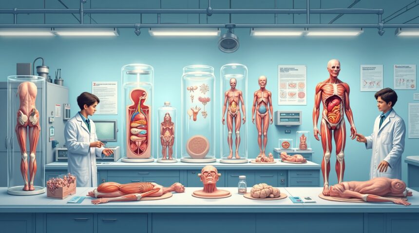
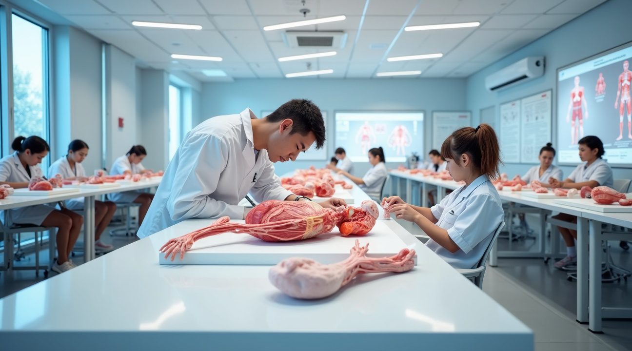
Medical schools worldwide now use plastinated specimens as primary teaching tools. Students examine internal structures without exposure to harmful chemicals. Professors demonstrate anatomical relationships that traditional preservation methods often obscure. Museums display plastinated bodies for public education, sparking both fascination and ethical debates.
Plastination Process Overview
- Fixation – Use of formaldehyde or similar agents to halt decay and initiate tissue stabilization.
- Dehydration – Specimens are submerged in cold acetone baths to remove water and soluble fats.
- Forced Impregnation – Vacuum chambers draw polymer mixtures into evacuated cells previously occupied by water and fats.
- Positioning – Specimens are posed anatomically while polymers remain pliable.
- Curing – Application of gas, heat, or UV light hardens polymers into durable forms.
- Final Quality Check – Each specimen is scrutinized for completeness, stability, and teaching value.
Different polymer systems serve specific educational purposes. Silicone rubber produces flexible specimens ideal for manipulation during teaching sessions. Epoxy resins create transparent sections that reveal internal structures clearly. Polyester compounds generate thin slices perfect for microscopic examination and detailed study.
Quality control measures ensure specimen integrity throughout processing. Temperature monitoring prevents tissue damage during dehydration phases. Vacuum levels require constant adjustment to achieve optimal polymer penetration. Processing times vary significantly based on specimen size and density.
Institutional Impact and Global Practices
Educational institutions invest substantially in plastination laboratories due to equipment costs and specialized training requirements. However, long-term benefits include reduced storage expenses and elimination of chemical replacement needs. Specimens last decades without deterioration, providing consistent educational resources for multiple student generations.
Ethical considerations shape every aspect of plastination programs. Donors must understand their bodies may be displayed publicly for extended periods. Families receive detailed information about processing procedures and final specimen uses. Institutions maintain strict documentation of consent and specimen tracking.
International regulations govern plastination practices across different countries. Some nations restrict public displays while others encourage educational exhibitions. Legal frameworks continue evolving as the technology gains acceptance in medical and educational communities worldwide.
For additional details on plastination, you can visit the official Body Worlds exhibition website to explore how this technology is used in real-world educational displays.

From Formaldehyde to Forever: How Dr. Gunther von Hagens Revolutionized Anatomical Preservation in 1977
I find it fascinating how one man’s innovation completely transformed the field of anatomical preservation. Dr. Gunther von Hagens introduced plastination in 1977, creating a revolutionary technique that replaced traditional formaldehyde-based preservation methods. His breakthrough fundamentally changed how scientists, educators, and medical professionals could study and display biological specimens.
The Plastination Process and Polymer Selection
Plastination works by systematically replacing all water and fats within biological tissues with specialized curable polymers. During this intricate process, specimens undergo several stages where natural fluids get substituted with synthetic materials that harden into permanent structures. The technique creates what scientists call plastinates—preserved specimens that maintain their original form while gaining remarkable durability.
Three primary polymers drive the plastination process, each serving specific purposes:
- Silicone rubber provides flexibility and maintains natural tissue texture, making it ideal for organs and soft tissue specimens
- Epoxy resin creates transparent, hard specimens perfect for demonstrating internal structures and cross-sections
- Polyester-copolymer offers excellent detail preservation for thin tissue slices and microscopic specimens
Revolutionary Advantages Over Traditional Methods
The brilliance of von Hagens’ invention lies in what plastinates don’t do. Unlike traditional preservation methods that rely on formaldehyde or other chemicals, plastinated specimens don’t emit unpleasant odors that can overwhelm laboratory spaces. More importantly, they don’t decompose or decay over time, eliminating the need for constant maintenance or replacement.
This durability factor has proven invaluable for educational institutions and medical facilities. Students can handle plastinated specimens directly without gloves or special ventilation systems. The specimens maintain their educational value indefinitely, making them cost-effective investments for long-term use.
I observe that plastination has opened doors for public education that were previously impossible. Museums now display human anatomy exhibits that engage visitors without the ethical and practical concerns associated with traditional preservation methods. The process has democratized anatomical education, bringing complex biological structures within reach of general audiences.
The transformation from von Hagens’ initial concept to today’s widespread application demonstrates how scientific innovation can bridge gaps between research, education, and public understanding. Modern plastination facilities now produce specimens for medical schools, research institutions, and educational centers worldwide, continuing the revolutionary impact that began with a single breakthrough in 1977.
To learn more about Dr. Gunther von Hagens and his work, you can visit the official BODY WORLDS website.
https://www.youtube.com/watch?v=gPvI_B_zSSI

The Six-Step Journey: Breaking Down the Complete Plastination Process
The plastination process transforms biological specimens into durable educational tools through a precise six-step procedure that can take up to ten months to complete. Each phase builds upon the previous one, requiring specialized equipment and expert knowledge to achieve the remarkable preservation that makes plastinated specimens so valuable for scientific study.
Initial Preparation and Structural Exposure
The journey begins with fixation, where I immerse the specimen in formaldehyde or other preservation solutions. This critical first step kills bacteria and halts decomposition, creating a stable foundation for the remaining processes. The fixation typically requires 3–4 hours, though timing varies based on specimen size and density.
Dissection follows as the most labor-intensive phase of the entire process. Skilled dissectors carefully remove skin, fat, and connective tissue to expose the anatomical structures that will become the focus of the final display. This painstaking work can consume up to 1,000 hours for complex specimens, as each cut must preserve the integrity of delicate structures while revealing the educational features that make the specimen valuable.
The third step involves dehydration and defatting, where specimens undergo immersion in cold acetone maintained at temperatures between -20°C and -30°C. The acetone volume must be ten times greater than the specimen itself, and I change this solution multiple times throughout the process. This phase removes water and soluble fats from tissues, preparing them for polymer infiltration. The process extends up to four months, as thorough dehydration proves essential for successful polymer penetration.
Polymer Integration and Final Transformation
Forced impregnation represents the technical heart of plastination. During this fourth step, I submerge the specimen in liquid polymer and place it under vacuum conditions. The vacuum causes acetone to vaporize while simultaneously drawing polymer solution into every cell and tissue space. This remarkable exchange typically requires about two months to complete, as the polymer must reach even the most microscopic cellular structures.
Positioning demands both artistic vision and anatomical expertise. While the specimen remains flexible from polymer saturation, I use wires, needles, and foam blocks to achieve precise anatomical alignment. This step transforms the preserved tissue into an educational tool that accurately represents natural positioning and relationships between structures. Expert anatomical knowledge becomes crucial during this 2.5-month phase, as improper positioning can compromise the specimen’s educational value.
The final curing process solidifies the polymer through gas exposure, heat application, or ultraviolet light treatment. This hardening phase typically spans 1.5 to 2 months, during which the flexible polymer transforms into the durable material that characterizes finished plastinated specimens. The curing method varies depending on the specific polymer used and the intended final properties of the specimen.
Each step in this journey requires precise timing and environmental control. Temperature, humidity, and chemical concentrations must remain within strict parameters throughout the process. The extended timeline reflects the thoroughness needed to achieve complete preservation while maintaining the structural integrity that makes plastinated specimens so effective for education and research. Much like how smart glasses work through complex technological processes, plastination relies on carefully orchestrated chemical and physical transformations to achieve its remarkable results.

The Laboratory Behind the Magic: Essential Equipment and Materials for Success
Setting up a plastination laboratory requires specialized equipment that can handle the demanding requirements of this preservation process. I’ve found that successful plastination depends heavily on having the right tools and maintaining precise environmental controls throughout the procedure.
Critical Equipment for Professional Plastination
The backbone of any plastination laboratory centers around several key pieces of equipment:
- Vacuum pumps capable of creating and maintaining strong suction for polymer impregnation
- Industrial freezers that can consistently maintain temperatures at -25°C
- High-quality vacuum tubing to connect various components of the system
- Glass jars in multiple sizes for specimen processing and storage
- Heavy-duty plastic bags designed to withstand chemical exposure and temperature fluctuations
Temperature control represents one of the most critical aspects of the entire process. Dehydration stages require maintaining temperatures between -20°C to -30°C, demanding freezers that can operate reliably within this narrow range. Any deviation from these specifications can compromise the final quality of the preserved specimen.
Beyond the major equipment, laboratories must stock essential consumables throughout the lengthy process:
- Polymers serve as the replacement medium for biological fluids
- Formalin acts as the primary fixative for initial preservation
- Acetone plays a crucial role during the dehydration phase, gradually replacing water within the tissues
The time investment for plastination proves substantial, requiring extraordinary patience and dedication. Processing a complete human body demands up to 1,000 hours of active laboratory work. This doesn’t include the passive time during which specimens undergo chemical processes without direct intervention.
The total duration for plastinating an entire body extends well over a year. Individual steps within the process consume considerable time – impregnation and curing phases alone can take several months to complete properly. Each stage cannot be rushed without risking the integrity of the final specimen.
Laboratory staff must understand that plastination operates on biological timelines rather than industrial schedules. Tissues need adequate time to absorb polymers completely, and any attempt to accelerate these natural processes typically results in inferior preservation quality.
The investment in proper laboratory equipment pays dividends in the consistency and quality of results. Substandard vacuum pumps may fail to achieve necessary pressure levels, while inadequate freezing capacity can lead to temperature fluctuations that damage specimens. Professional-grade equipment ensures reliable operation during the extended processing periods that plastination requires.
Space and Storage Considerations
Storage capacity becomes another important consideration when planning laboratory space. Multiple specimens at different stages of processing require dedicated storage areas, each maintained at specific temperatures and conditions. Glass jars must be available in various sizes to accommodate different anatomical structures and processing requirements.
Safety and Chemical Handling
Chemical handling protocols demand careful attention to safety and proper ventilation systems. Acetone, formalin, and various polymers require specialized storage and handling procedures to protect laboratory personnel and maintain chemical integrity. Proper ventilation prevents accumulation of potentially harmful vapors during extended processing periods.
Human Resources and Monitoring
The labor-intensive nature of plastination means that even small-scale operations require significant human resources. Monitoring specimens, adjusting equipment settings, and performing regular maintenance tasks consume substantial time throughout the year-long process. Successful laboratories often employ multiple technicians to ensure continuous coverage during critical processing phases.
Quality Control and Redundancy
Quality control measures become essential when managing such lengthy procedures. Regular monitoring of temperatures, vacuum levels, and chemical concentrations prevents costly mistakes that could ruin months of work. Documentation of each processing step allows laboratories to troubleshoot problems and refine their techniques over time.
Investment in backup systems proves wise given the extended timelines involved. Redundant freezers, backup vacuum pumps, and emergency power systems protect valuable specimens from equipment failures that could occur during the months-long processing cycle.
For an example of plastination in practice, this video provides additional insight:
https://www.youtube.com/watch?v=wSxKQo4X2QY

Three Pathways to Perfection: Understanding Different Types of Plastinated Specimens
The plastination process employs distinct polymer systems, each engineered for specific applications and educational goals. These specialized techniques transform preserved specimens into teaching tools that serve different purposes in medical education and anatomical study.
Flexible Solutions: Silicone-Based Specimens
Silicone impregnated specimens represent the most versatile category in plastination technology. This method creates flexible, durable anatomical models that maintain their natural texture while offering exceptional longevity. Medical schools and training facilities favor these specimens because students can manipulate them repeatedly without causing damage. The silicone polymer penetrates every cellular space, replacing water and fat to create specimens that retain their three-dimensional structure indefinitely.
These silicone specimens excel at demonstrating anatomical relationships between organs, muscles, and skeletal structures. Unlike rigid displays, educators can bend joints, separate muscle layers, and show how organs connect within body cavities. The flexibility allows for dynamic teaching sessions where instructors demonstrate movement patterns and spatial relationships that static models cannot provide.
Clarity and Precision: Specialized Polymer Applications
The Polyester P40 technique opens up entirely different educational possibilities through its ability to create transparent sections. This method proves particularly valuable for brain slices and thin tissue sheets where internal structures need clear visualization. Neuroanatomy courses benefit significantly from P40 specimens because students can observe neural pathways, gray matter distribution, and cellular arrangements without traditional sectioning limitations.
Sheet plastinates created through this process offer remarkable detail for studying organ architecture. These transparent specimens allow examination of:
- Vascular networks within organs
- Tissue layer organization
- Cellular distribution patterns
- Anatomical boundaries between structures
- Microscopic detail preservation
Epoxy resin provides the ultimate in rigidity and clarity for specialized applications requiring maximum structural integrity. These crystal-clear specimens work exceptionally well for detailed bone studies, joint mechanics demonstrations, and permanent museum displays. The epoxy system creates specimens so transparent that internal features become visible without compromising structural strength.
Enhanced visualization reaches new levels through latex injection techniques that highlight circulatory systems. Technicians inject red latex into arterial vessels and blue latex into venous systems before beginning the plastination process. This color-coding system transforms complex vascular networks into easily identifiable educational tools. Students can trace blood flow patterns, understand circulation pathways, and grasp cardiovascular anatomy concepts that would otherwise require extensive dissection.
The latex injection process requires precise timing and pressure control to ensure complete vessel filling without rupturing delicate capillary networks. Once the colored latex sets, the subsequent plastination process preserves both the specimen structure and the enhanced vascular visualization permanently.
Each polymer system serves distinct educational objectives while maintaining the fundamental plastination principle of cellular replacement. Silicone specimens provide hands-on flexibility for interactive learning, P40 creates transparent windows into anatomical structures, epoxy delivers maximum clarity for detailed observation, and latex injection enhances vascular comprehension. These varied approaches ensure that plastination technology meets diverse educational needs across medical training programs, research facilities, and public education initiatives.
The selection between these pathways depends on intended use, required durability, and specific anatomical features being emphasized. Modern plastination facilities often employ multiple techniques simultaneously to create comprehensive specimen collections that address different learning styles and educational requirements.

Why Plastination Beats Traditional Methods: The Educational Revolution in Medical Training
Medical education has undergone a dramatic transformation with the introduction of plastination technology. This preservation method offers significant advantages over traditional wet-preserved cadavers and other conventional techniques, fundamentally changing how anatomy is taught in classrooms worldwide.
Plastinates deliver superior anatomical visualization compared to formaldehyde-preserved specimens, which often appear discolored and deteriorated over time. The plastic polymer replacement maintains natural colors and textures, allowing students to observe anatomical structures with remarkable clarity. Unlike formalin-preserved specimens that become increasingly difficult to examine as they age, plastinated materials retain their educational value for decades.
Practical Benefits for Educational Institutions
The durability and portability of plastinated specimens address many logistical challenges faced by medical schools. Consider these key advantages that make plastination ideal for modern anatomy teaching:
- No special storage requirements – specimens can be kept at room temperature without refrigeration
- Complete elimination of toxic formaldehyde odors that often overwhelm traditional dissection laboratories
- Safe handling without protective gloves or ventilation concerns
- Exceptional long-term preservation lasting many years without decay
- Lightweight construction allowing easy transport between classrooms and institutions
- Non-toxic composition ensuring student and faculty safety
These practical benefits enable institutions to expand their anatomy programs beyond traditional laboratory settings. Plastinated specimens can be used in regular classrooms, field studies, or even remote learning environments where conventional preservation methods would be impractical.
Despite these advantages, plastination does present certain limitations. The process requires considerable time and skilled technicians to complete successfully. Additionally, plastinated specimens become rigid during processing, which limits their ability to demonstrate joint movement and flexibility that living tissue possesses. Students also miss the tactile experience of working with wet tissue, and the specimens cannot be dissected for hands-on surgical practice.
Cryopreservation and traditional formalin preservation remain competitive alternatives, each offering distinct benefits. However, for long-term educational use and classroom safety, plastination has established itself as the premier preservation method. The technology continues to revolutionize medical training by providing institutions with durable, safe, and educationally superior anatomical specimens that enhance student learning while reducing operational costs and safety concerns.

The Human Side of Science: Examining Ethical and Legal Boundaries in Body Donation
Body donation for plastination involves strict legal and ethical protocols that protect both donors and their families. I’ve observed how institutions must follow comprehensive guidelines that govern every aspect of the donation process, from initial consent through final display. The legal framework varies by jurisdiction, but most require written consent that specifically addresses long-term preservation and potential public exhibition of plastinated specimens.
Explicit donor consent becomes particularly critical when specimens will be displayed publicly or used for extended periods. Donors must understand that plastination creates permanent specimens that could be viewed by thousands of people over many years. Legal documents typically outline specific uses, including educational demonstrations, museum exhibitions, and research applications. Family members often retain certain rights regarding the eventual disposition of plastinated remains, even after the initial donation agreement.
Educational and Exhibition Applications
Plastinated specimens serve dual purposes in education and public exhibition, each requiring distinct ethical considerations. Educational institutions use these preserved bodies to teach anatomy, physiology, and medical procedures to students and healthcare professionals. Museums and science centers display plastinates to educate the general public about human anatomy and health. However, both contexts demand careful adherence to established guidelines that respect human dignity while maximizing educational value.
Professional organizations have developed specific protocols for displaying human specimens. These guidelines address everything from lighting and signage to visitor interaction policies. Educational facilities must ensure that plastinates are presented respectfully, with appropriate context and scientific information. Public exhibitions often include detailed explanations about the donation process and the scientific significance of plastination, helping visitors understand the contribution made by donors.
Labor-Intensive Preparation Process
The transformation of donated bodies into educational specimens requires extraordinary time and skill investment. Specimen preparation typically demands between 500 to 1,000 hours of specialized labor, depending on the complexity of the final display. This extensive timeline reflects the precision required at each stage, from initial dissection through final positioning and curing.
Skilled personnel must possess both anatomical knowledge and technical expertise in plastination procedures. The process involves several specialized roles:
- Anatomical dissection specialists who carefully expose specific body systems
- Plastination technicians who manage the polymer replacement process
- Positioning experts who arrange specimens for optimal educational impact
- Quality control specialists who ensure preservation standards are met
Each step requires careful documentation and quality assurance measures. Personnel must maintain detailed records of procedures, materials used, and preservation conditions. This documentation serves multiple purposes, including legal compliance, quality control, and educational reference materials for institutions using the specimens.
Professional training programs have emerged to address the growing demand for qualified plastination technicians. These programs combine anatomical study with hands-on experience in polymer chemistry and preservation techniques. The specialized nature of this work means that skilled practitioners often command significant compensation, reflecting both their expertise and the critical role they play in advancing medical education.
The ethical dimensions extend beyond donor consent to include fair labor practices and appropriate recognition for the skilled professionals who make plastination possible. Institutions must balance the significant costs associated with quality specimen preparation against their educational mission and budget constraints. This balance often influences decisions about which specimens to create and how extensively they’ll be used in educational programs.

Sources: